6B25

Crystal structure of human STAC1 Tandem SH3 Domains (288-402)


Experimental Data Snapshot

  • Method: X-RAY DIFFRACTION
  • Resolution: 2.39 Å
  • R-Value Free: 0.259 
  • R-Value Work: 0.203 
  • R-Value Observed: 0.205 

wwPDB Validation   3D Report Full Report


This is version 1.5 of the entry. See complete history


Literature

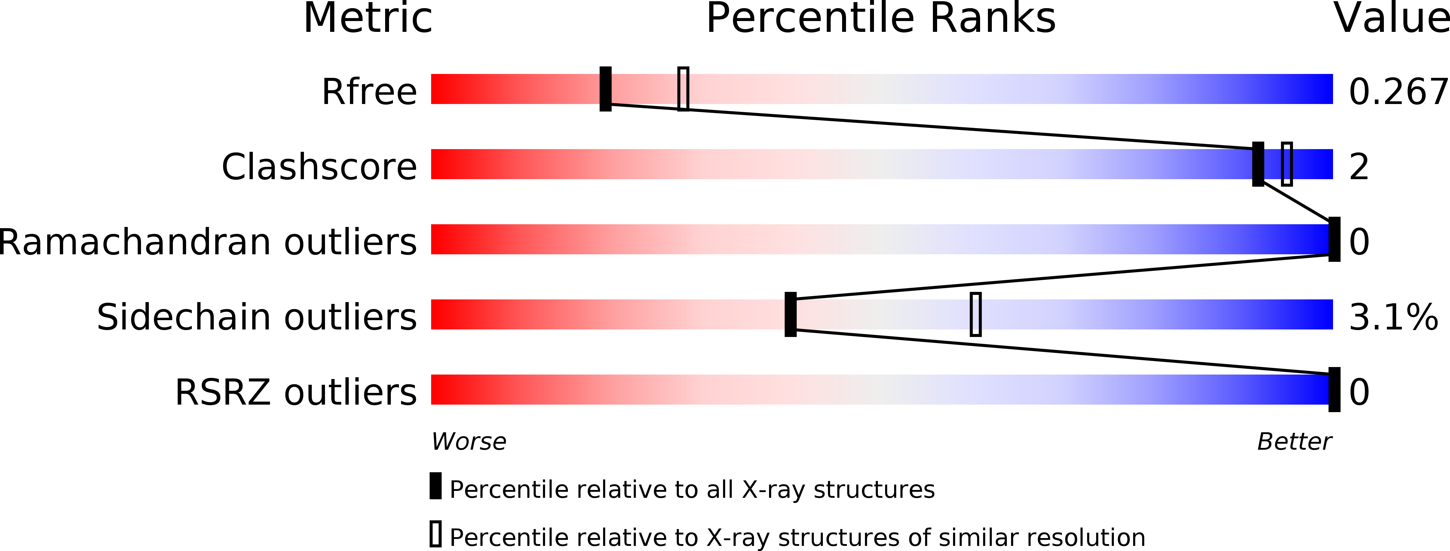
Structural insights into binding of STAC proteins to voltage-gated calcium channels.

Wong King Yuen, S.M.Campiglio, M.Tung, C.C.Flucher, B.E.Van Petegem, F.

(2017) Proc Natl Acad Sci U S A 114: E9520-E9528

  • DOI: https://doi.org/10.1073/pnas.1708852114
  • Primary Citation of Related Structures:  
    6B25, 6B26, 6B27, 6B28, 6B29

  • PubMed Abstract: 

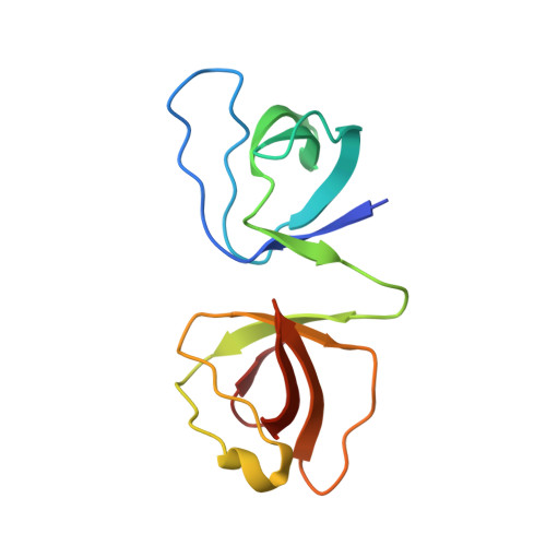
    Excitation-contraction (EC) coupling in skeletal muscle requires functional and mechanical coupling between L-type voltage-gated calcium channels (Ca V 1.1) and the ryanodine receptor (RyR1). Recently, STAC3 was identified as an essential protein for EC coupling and is part of a group of three proteins that can bind and modulate L-type voltage-gated calcium channels. Here, we report crystal structures of tandem-SH3 domains of different STAC isoforms up to 1.2-Å resolution. These form a rigid interaction through a conserved interdomain interface. We identify the linker connecting transmembrane repeats II and III in two different Ca V isoforms as a binding site for the SH3 domains and report a crystal structure of the complex with the STAC2 isoform. The interaction site includes the location for a disease variant in STAC3 that has been linked to Native American myopathy (NAM). Introducing the mutation does not cause misfolding of the SH3 domains, but abolishes the interaction. Disruption of the interaction via mutations in the II-III loop perturbs skeletal muscle EC coupling, but preserves the ability of STAC3 to slow down inactivation of Ca V 1.2.


  • Organizational Affiliation

    Department of Biochemistry and Molecular Biology, University of British Columbia, V6T 1Z3 Vancouver, BC, Canada.


Macromolecules
Find similar proteins by:  (by identity cutoff)  |  3D Structure
Entity ID: 1
MoleculeChains Sequence LengthOrganismDetailsImage
SH3 and cysteine-rich domain-containing protein118Homo sapiensMutation(s): 0 
Gene Names: STAC
UniProt & NIH Common Fund Data Resources
Find proteins for Q99469 (Homo sapiens)
Explore Q99469 
Go to UniProtKB:  Q99469
PHAROS:  Q99469
GTEx:  ENSG00000144681 
Entity Groups  
Sequence Clusters30% Identity50% Identity70% Identity90% Identity95% Identity100% Identity
UniProt GroupQ99469
Sequence Annotations
Expand
  • Reference Sequence
Small Molecules
Ligands 1 Unique
IDChains Name / Formula / InChI Key2D Diagram3D Interactions
EPE
Query on EPE

Download Ideal Coordinates CCD File 
B [auth A]4-(2-HYDROXYETHYL)-1-PIPERAZINE ETHANESULFONIC ACID
C8 H18 N2 O4 S
JKMHFZQWWAIEOD-UHFFFAOYSA-N
Experimental Data & Validation

Experimental Data

  • Method: X-RAY DIFFRACTION
  • Resolution: 2.39 Å
  • R-Value Free: 0.259 
  • R-Value Work: 0.203 
  • R-Value Observed: 0.205 
  • Space Group: P 21 21 21
Unit Cell:
Length ( Å )Angle ( ˚ )
a = 28.958α = 90
b = 36.727β = 90
c = 113.001γ = 90
Software Package:
Software NamePurpose
HKL-2000data collection
SCALEPACKdata scaling
REFMACrefinement
PDB_EXTRACTdata extraction
DENZOdata reduction
REFMACphasing

Structure Validation

View Full Validation Report


Entry History & Funding Information

Deposition Data


Funding OrganizationLocationGrant Number
Canadian Institutes of Health Research (CIHR)CanadaMOP-119608

Revision History  (Full details and data files)

  • Version 1.0: 2017-10-25
    Type: Initial release
  • Version 1.1: 2017-11-08
    Changes: Database references
  • Version 1.2: 2017-11-22
    Changes: Database references
  • Version 1.3: 2017-12-06
    Changes: Author supporting evidence
  • Version 1.4: 2020-01-08
    Changes: Author supporting evidence
  • Version 1.5: 2024-03-13
    Changes: Data collection, Database references