The C-terminal domain of the MutL homolog from Neisseria gonorrhoeae forms an inverted homodimer
Namadurai, S., Jain, D., Kulkarni, D.S., Tabib, C.R., Friedhoff, P., Rao, D.N., Nair, D.T.(2010) PLoS One 5: e13726-e13726
- PubMed: 21060849
- DOI: https://doi.org/10.1371/journal.pone.0013726
- Primary Citation of Related Structures:
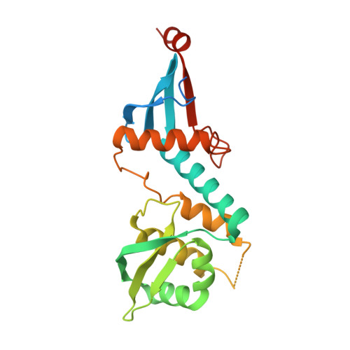
3NCV - PubMed Abstract:
The mismatch repair (MMR) pathway serves to maintain the integrity of the genome by removing mispaired bases from the newly synthesized strand. In E. coli, MutS, MutL and MutH coordinate to discriminate the daughter strand through a mechanism involving lack of methylation on the new strand. This facilitates the creation of a nick by MutH in the daughter strand to initiate mismatch repair. Many bacteria and eukaryotes, including humans, do not possess a homolog of MutH. Although the exact strategy for strand discrimination in these organisms is yet to be ascertained, the required nicking endonuclease activity is resident in the C-terminal domain of MutL. This activity is dependent on the integrity of a conserved metal binding motif. Unlike their eukaryotic counterparts, MutL in bacteria like Neisseria exist in the form of a homodimer. Even though this homodimer would possess two active sites, it still acts a nicking endonuclease. Here, we present the crystal structure of the C-terminal domain (CTD) of the MutL homolog of Neisseria gonorrhoeae (NgoL) determined to a resolution of 2.4 Å. The structure shows that the metal binding motif exists in a helical configuration and that four of the six conserved motifs in the MutL family, including the metal binding site, localize together to form a composite active site. NgoL-CTD exists in the form of an elongated inverted homodimer stabilized by a hydrophobic interface rich in leucines. The inverted arrangement places the two composite active sites in each subunit on opposite lateral sides of the homodimer. Such an arrangement raises the possibility that one of the active sites is occluded due to interaction of NgoL with other protein factors involved in MMR. The presentation of only one active site to substrate DNA will ensure that nicking of only one strand occurs to prevent inadvertent and deleterious double stranded cleavage.
Organizational Affiliation:
Laboratory 4, National Centre for Biological Sciences, Bangalore, India.