2XJZ

Crystal structure of the LMO2:LDB1-LID complex, C2 crystal form


Experimental Data Snapshot

  • Method: X-RAY DIFFRACTION
  • Resolution: 2.80 Å
  • R-Value Free: 0.244 
  • R-Value Work: 0.228 
  • R-Value Observed: 0.229 

wwPDB Validation   3D Report Full Report


This is version 1.3 of the entry. See complete history


Literature

Structure of the Leukemia Oncogene Lmo2: Implications for the Assembly of a Hematopoietic Transcription Factor Complex.

El Omari, K.Hoosdally, S.J.Tuladhar, K.Karia, D.Vyas, P.Patient, R.Porcher, C.Mancini, E.J.

(2011) Blood 117: 2146

  • DOI: https://doi.org/10.1182/blood-2010-07-293357
  • Primary Citation of Related Structures:  
    2XJY, 2XJZ

  • PubMed Abstract: 

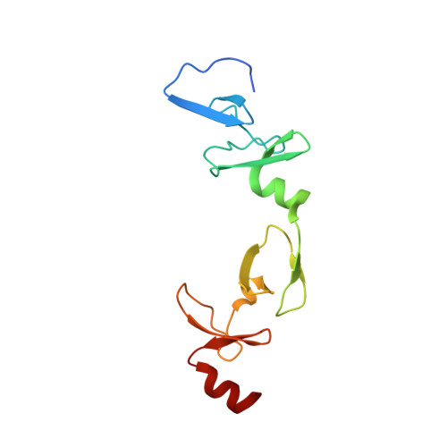
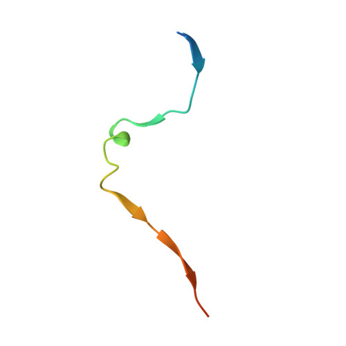
    The LIM only protein 2 (LMO2) is a key regulator of hematopoietic stem cell development whose ectopic expression in T cells leads to the onset of acute lymphoblastic leukemia. Through its LIM domains, LMO2 is thought to function as the scaffold for a DNA-binding transcription regulator complex, including the basic helix-loop-helix proteins SCL/TAL1 and E47, the zinc finger protein GATA-1, and LIM-domain interacting protein LDB1. To understand the role of LMO2 in the formation of this complex and ultimately to dissect its function in normal and aberrant hematopoiesis, we solved the crystal structure of LMO2 in complex with the LID domain of LDB1 at 2.4 Å resolution. We observe a largely unstructured LMO2 kept in register by the LID binding both LIM domains. Comparison of independently determined crystal structures of LMO2 reveals large movements around a conserved hinge between the LIM domains. We demonstrate that such conformational flexibility is necessary for binding of LMO2 to its partner protein SCL/TAL1 in vitro and for the function of this complex in vivo. These results, together with molecular docking and analysis of evolutionarily conserved residues, yield the first structural model of the DNA-binding complex containing LMO2, LDB1, SCL/TAL1, and GATA-1.


  • Organizational Affiliation

    Division of Structural Biology, Wellcome Trust Centre for Human Genetics, University of Oxford, Oxford, UK.


Macromolecules
Find similar proteins by:  (by identity cutoff)  |  3D Structure
Entity ID: 1
MoleculeChains Sequence LengthOrganismDetailsImage
RHOMBOTIN-2
A, B, C, D, E
131Homo sapiensMutation(s): 0 
UniProt & NIH Common Fund Data Resources
Find proteins for P25791 (Homo sapiens)
Explore P25791 
Go to UniProtKB:  P25791
PHAROS:  P25791
GTEx:  ENSG00000135363 
Entity Groups  
Sequence Clusters30% Identity50% Identity70% Identity90% Identity95% Identity100% Identity
UniProt GroupP25791
Sequence Annotations
Expand
  • Reference Sequence
Find similar proteins by:  (by identity cutoff)  |  3D Structure
Entity ID: 2
MoleculeChains Sequence LengthOrganismDetailsImage
LIM DOMAIN-BINDING PROTEIN 1F [auth I],
G [auth J],
H [auth K],
I [auth L],
J [auth M]
36Homo sapiensMutation(s): 0 
UniProt & NIH Common Fund Data Resources
Find proteins for Q86U70 (Homo sapiens)
Explore Q86U70 
Go to UniProtKB:  Q86U70
PHAROS:  Q86U70
GTEx:  ENSG00000198728 
Entity Groups  
Sequence Clusters30% Identity50% Identity70% Identity90% Identity95% Identity100% Identity
UniProt GroupQ86U70
Sequence Annotations
Expand
  • Reference Sequence
Small Molecules
Ligands 2 Unique
IDChains Name / Formula / InChI Key2D Diagram3D Interactions
ZN
Query on ZN

Download Ideal Coordinates CCD File 
AA [auth D]
CA [auth E]
DA [auth E]
EA [auth E]
FA [auth E]
AA [auth D],
CA [auth E],
DA [auth E],
EA [auth E],
FA [auth E],
K [auth A],
L [auth A],
M [auth A],
N [auth A],
O [auth B],
P [auth B],
Q [auth B],
R [auth B],
T [auth C],
U [auth C],
V [auth C],
W [auth C],
X [auth D],
Y [auth D],
Z [auth D]
ZINC ION
Zn
PTFCDOFLOPIGGS-UHFFFAOYSA-N
CL
Query on CL

Download Ideal Coordinates CCD File 
BA [auth D],
S [auth B]
CHLORIDE ION
Cl
VEXZGXHMUGYJMC-UHFFFAOYSA-M
Experimental Data & Validation

Experimental Data

  • Method: X-RAY DIFFRACTION
  • Resolution: 2.80 Å
  • R-Value Free: 0.244 
  • R-Value Work: 0.228 
  • R-Value Observed: 0.229 
  • Space Group: C 1 2 1
Unit Cell:
Length ( Å )Angle ( ˚ )
a = 179.92α = 90
b = 55.53β = 90.11
c = 114.71γ = 90
Software Package:
Software NamePurpose
BUSTERrefinement
HKL-2000data reduction
HKL-2000data scaling
PHENIXphasing

Structure Validation

View Full Validation Report


Entry History 

Deposition Data

Revision History  (Full details and data files)

  • Version 1.0: 2010-07-21
    Type: Initial release
  • Version 1.1: 2011-05-19
    Changes: Version format compliance
  • Version 1.2: 2011-07-13
    Changes: Version format compliance
  • Version 1.3: 2017-07-12
    Changes: Refinement description