7ZPP

Cryo-EM structure of the MVV CSC intasome at 4.5A resolution


Experimental Data Snapshot

  • Method: ELECTRON MICROSCOPY
  • Resolution: 4.50 Å
  • Aggregation State: PARTICLE 
  • Reconstruction Method: SINGLE PARTICLE 

wwPDB Validation   3D Report Full Report


This is version 1.2 of the entry. See complete history


Literature

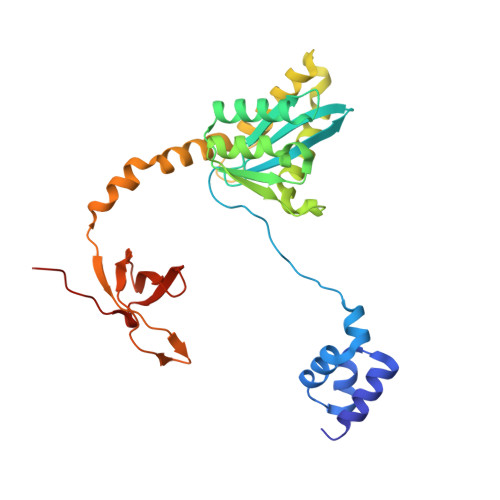
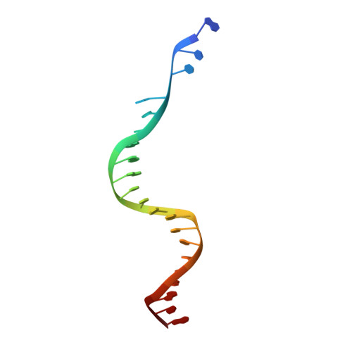
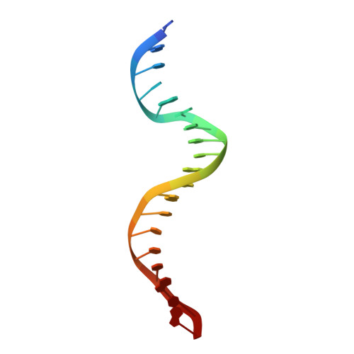
A supramolecular assembly mediates lentiviral DNA integration

Ballandras-Colas, A.Maskell, D.P.Serrao, E.Locke, J.Swuec, P.Jonsson, S.R.Kotecha, A.Cook, N.J.Pye, V.E.Taylor, I.A.Andresdottir, V.Engelman, A.N.Costa, A.Cherepanov, P.

(2017) Science 355: 93-95

  • DOI: https://doi.org/10.1126/science.aah7002
  • Primary Citation of Related Structures:  
    5LLJ, 5M0R, 5T3A, 7ZPP

  • PubMed Abstract: 

    Retroviral integrase (IN) functions within the intasome nucleoprotein complex to catalyze insertion of viral DNA into cellular chromatin. Using cryo-electron microscopy, we now visualize the functional maedi-visna lentivirus intasome at 4.9 angstrom resolution. The intasome comprises a homo-hexadecamer of IN with a tetramer-of-tetramers architecture featuring eight structurally distinct types of IN protomers supporting two catalytically competent subunits. The conserved intasomal core, previously observed in simpler retroviral systems, is formed between two IN tetramers, with a pair of C-terminal domains from flanking tetramers completing the synaptic interface. Our results explain how HIV-1 IN, which self-associates into higher-order multimers, can form a functional intasome, reconcile the bulk of early HIV-1 IN biochemical and structural data, and provide a lentiviral platform for design of HIV-1 IN inhibitors.


  • Organizational Affiliation

    Chromatin Structure and Mobile DNA, The Francis Crick Institute, London, NW1 1AT, UK.


Macromolecules

Find similar proteins by:  (by identity cutoff)  |  3D Structure
Entity ID: 1
MoleculeChains Sequence LengthOrganismDetailsImage
Integrase
A, B, C, D, E
A, B, C, D, E, F, G, H, I, J, K, L, M, N, O, P
281Visna/maedi virus EV1 KV1772Mutation(s): 0 
Gene Names: pol
EC: 2.7.7 (PDB Primary Data), 3.1 (PDB Primary Data)
UniProt
Find proteins for P35956 (Maedi visna virus (strain KV1772))
Explore P35956 
Go to UniProtKB:  P35956
Entity Groups  
Sequence Clusters30% Identity50% Identity70% Identity90% Identity95% Identity100% Identity
UniProt GroupP35956
Sequence Annotations
Expand
  • Reference Sequence

Find similar nucleic acids by:  Sequence   |   3D Structure  

Entity ID: 2
MoleculeChains LengthOrganismImage
vDNA, non-transferred strand
Q, S
21Visna lentivirus (strain 1514)
Sequence Annotations
Expand
  • Reference Sequence

Find similar nucleic acids by:  Sequence   |   3D Structure  

Entity ID: 3
MoleculeChains LengthOrganismImage
vDNA, transferred strand
R, T
19Visna-maedi virus
Sequence Annotations
Expand
  • Reference Sequence
Experimental Data & Validation

Experimental Data

  • Method: ELECTRON MICROSCOPY
  • Resolution: 4.50 Å
  • Aggregation State: PARTICLE 
  • Reconstruction Method: SINGLE PARTICLE 
EM Software:
TaskSoftware PackageVersion
MODEL REFINEMENTPHENIX
MODEL REFINEMENTCoot
RECONSTRUCTIONcryoSPARC2

Structure Validation

View Full Validation Report


Entry History & Funding Information

Deposition Data


Funding OrganizationLocationGrant Number
The Francis Crick InstituteUnited KingdomFC001061
Wellcome TrustUnited KingdomFC001061
Medical Research Council (MRC, United Kingdom)United KingdomFC001061
Cancer Research UKUnited KingdomFC001061

Revision History  (Full details and data files)

  • Version 1.0: 2022-05-11
    Type: Initial release
  • Version 1.1: 2022-06-29
    Changes: Advisory, Database references, Source and taxonomy, Structure summary
  • Version 1.2: 2022-10-12
    Changes: Structure summary