7WAW

MurJ inward closed form


Experimental Data Snapshot

  • Method: X-RAY DIFFRACTION
  • Resolution: 2.80 Å
  • R-Value Free: 0.273 
  • R-Value Work: 0.216 
  • R-Value Observed: 0.222 

wwPDB Validation   3D Report Full Report


Ligand Structure Quality Assessment 


This is version 1.2 of the entry. See complete history


Literature

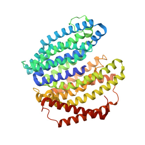
Crystal structure of the lipid flippase MurJ in a "squeezed" form distinct from its inward- and outward-facing forms.

Kohga, H.Mori, T.Tanaka, Y.Yoshikaie, K.Taniguchi, K.Fujimoto, K.Fritz, L.Schneider, T.Tsukazaki, T.

(2022) Structure 30: 1088-1097.e3

  • DOI: https://doi.org/10.1016/j.str.2022.05.008
  • Primary Citation of Related Structures:  
    7WAG, 7WAW, 7WAX

  • PubMed Abstract: 

    The bacterial peptidoglycan enclosing the cytoplasmic membrane is a fundamental cellular architecture. The integral membrane protein MurJ plays an essential role in flipping the cell wall building block Lipid II across the cytoplasmic membrane for peptidoglycan biosynthesis. Previously reported crystal structures of MurJ have elucidated its V-shaped inward- or outward-facing forms with an internal cavity for substrate binding. MurJ transports Lipid II using its cavity through conformational transitions between these two forms. Here, we report two crystal structures of inward-facing forms from Arsenophonus endosymbiont MurJ and an unprecedented crystal structure of Escherichia coli MurJ in a "squeezed" form, which lacks a cavity to accommodate the substrate, mainly because of the increased proximity of transmembrane helices 2 and 8. Subsequent molecular dynamics simulations supported the hypothesis that the squeezed form is an intermediate conformation. This study fills a gap in our understanding of the Lipid II flipping mechanism.


  • Organizational Affiliation

    Nara Institute of Science and Technology, Ikoma, Nara 630-0192, Japan.


Macromolecules
Find similar proteins by:  (by identity cutoff)  |  3D Structure
Entity ID: 1
MoleculeChains Sequence LengthOrganismDetailsImage
lipid II flippase MurJ511Candidatus Arsenophonus nilaparvataeMutation(s): 0 
Entity Groups  
Sequence Clusters30% Identity50% Identity70% Identity90% Identity95% Identity100% Identity
Sequence Annotations
Expand
  • Reference Sequence
Experimental Data & Validation

Experimental Data

  • Method: X-RAY DIFFRACTION
  • Resolution: 2.80 Å
  • R-Value Free: 0.273 
  • R-Value Work: 0.216 
  • R-Value Observed: 0.222 
  • Space Group: P 1 21 1
Unit Cell:
Length ( Å )Angle ( ˚ )
a = 56.41α = 90
b = 71.46β = 96.98
c = 74.54γ = 90
Software Package:
Software NamePurpose
PHENIXrefinement

Structure Validation

View Full Validation Report


Ligand Structure Quality Assessment 


Entry History & Funding Information

Deposition Data


Funding OrganizationLocationGrant Number
Japan Society for the Promotion of Science (JSPS)JapanJP21H05155
Japan Society for the Promotion of Science (JSPS)JapanJP21H05153
Japan Society for the Promotion of Science (JSPS)Japan21K19226
Japan Society for the Promotion of Science (JSPS)Japan21KK0125
Japan Society for the Promotion of Science (JSPS)Japan19K22395
Japan Society for the Promotion of Science (JSPS)Japan18KK0197
Japan Society for the Promotion of Science (JSPS)Japan18H02405

Revision History  (Full details and data files)

  • Version 1.0: 2022-06-01
    Type: Initial release
  • Version 1.1: 2022-06-22
    Changes: Database references
  • Version 1.2: 2022-08-17
    Changes: Database references