7SQ2

Reprocessed and refined structure of Phospholipase C-beta and Gq signaling complex


Experimental Data Snapshot

  • Method: X-RAY DIFFRACTION
  • Resolution: 2.60 Å
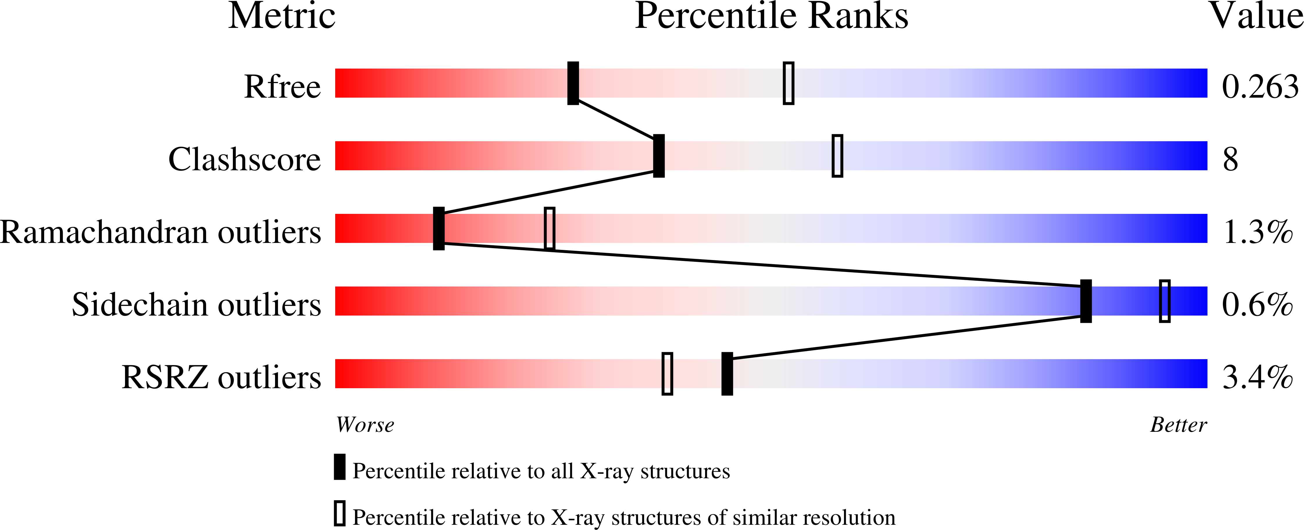
  • R-Value Free: 0.263 
  • R-Value Work: 0.199 
  • R-Value Observed: 0.202 

wwPDB Validation   3D Report Full Report


Ligand Structure Quality Assessment 


This is version 1.1 of the entry. See complete history

Re-refinement Note

This entry reflects an alternative modeling of the original data in: 3OHM


Literature

Kinetic Scaffolding Mediated by a Phospholipase C-{beta} and Gq Signaling Complex

Endo-Streeter, S.T.Sondek, J.Harden, T.K.

(2010) Science 330: 974-980

  • DOI: https://doi.org/10.1126/science.1193438
  • Primary Citation of Related Structures:  
    7SQ2

  • PubMed Abstract: 

    Transmembrane signals initiated by a broad range of extracellular stimuli converge on nodes that regulate phospholipase C (PLC)-dependent inositol lipid hydrolysis for signal propagation. We describe how heterotrimeric guanine nucleotide-binding proteins (G proteins) activate PLC-βs and in turn are deactivated by these downstream effectors. The 2.7-angstrom structure of PLC-β3 bound to activated Gα(q) reveals a conserved module found within PLC-βs and other effectors optimized for rapid engagement of activated G proteins. The active site of PLC-β3 in the complex is occluded by an intramolecular plug that is likely removed upon G protein-dependent anchoring and orientation of the lipase at membrane surfaces. A second domain of PLC-β3 subsequently accelerates guanosine triphosphate hydrolysis by Gα(q), causing the complex to dissociate and terminate signal propagation. Mutations within this domain dramatically delay signal termination in vitro and in vivo. Consequently, this work suggests a dynamic catch-and-release mechanism used to sharpen spatiotemporal signals mediated by diverse sensory inputs.


  • Organizational Affiliation

    Department of Pharmacology, University of North Carolina School of Medicine, Chapel Hill, NC 27599, USA.


Macromolecules
Find similar proteins by:  (by identity cutoff)  |  3D Structure
Entity ID: 1
MoleculeChains Sequence LengthOrganismDetailsImage
Guanine nucleotide-binding protein G(q) subunit alpha327Mus musculusMutation(s): 0 
Gene Names: Gnaq
UniProt
Find proteins for P21279 (Mus musculus)
Explore P21279 
Go to UniProtKB:  P21279
Entity Groups  
Sequence Clusters30% Identity50% Identity70% Identity90% Identity95% Identity100% Identity
UniProt GroupP21279
Sequence Annotations
Expand
  • Reference Sequence
Find similar proteins by:  (by identity cutoff)  |  3D Structure
Entity ID: 2
MoleculeChains Sequence LengthOrganismDetailsImage
1-phosphatidylinositol 4,5-bisphosphate phosphodiesterase beta-3885Homo sapiensMutation(s): 0 
Gene Names: PLCB3
EC: 3.1.4.11
UniProt & NIH Common Fund Data Resources
Find proteins for Q01970 (Homo sapiens)
Explore Q01970 
Go to UniProtKB:  Q01970
PHAROS:  Q01970
GTEx:  ENSG00000149782 
Entity Groups  
Sequence Clusters30% Identity50% Identity70% Identity90% Identity95% Identity100% Identity
UniProt GroupQ01970
Sequence Annotations
Expand
  • Reference Sequence
Small Molecules
Ligands 5 Unique
IDChains Name / Formula / InChI Key2D Diagram3D Interactions
GDP (Subject of Investigation/LOI)
Query on GDP

Download Ideal Coordinates CCD File 
E [auth A]GUANOSINE-5'-DIPHOSPHATE
C10 H15 N5 O11 P2
QGWNDRXFNXRZMB-UUOKFMHZSA-N
ALF (Subject of Investigation/LOI)
Query on ALF

Download Ideal Coordinates CCD File 
C [auth A]TETRAFLUOROALUMINATE ION
Al F4
UYOMQIYKOOHAMK-UHFFFAOYSA-J
ACT
Query on ACT

Download Ideal Coordinates CCD File 
F [auth A]
G [auth B]
H [auth B]
I [auth B]
J [auth B]
F [auth A],
G [auth B],
H [auth B],
I [auth B],
J [auth B],
K [auth B],
L [auth B]
ACETATE ION
C2 H3 O2
QTBSBXVTEAMEQO-UHFFFAOYSA-M
CA (Subject of Investigation/LOI)
Query on CA

Download Ideal Coordinates CCD File 
M [auth B]CALCIUM ION
Ca
BHPQYMZQTOCNFJ-UHFFFAOYSA-N
MG (Subject of Investigation/LOI)
Query on MG

Download Ideal Coordinates CCD File 
D [auth A],
N [auth B]
MAGNESIUM ION
Mg
JLVVSXFLKOJNIY-UHFFFAOYSA-N
Experimental Data & Validation

Experimental Data

  • Method: X-RAY DIFFRACTION
  • Resolution: 2.60 Å
  • R-Value Free: 0.263 
  • R-Value Work: 0.199 
  • R-Value Observed: 0.202 
  • Space Group: C 1 2 1
Unit Cell:
Length ( Å )Angle ( ˚ )
a = 202.837α = 90
b = 90.817β = 101.19
c = 93.135γ = 90
Software Package:
Software NamePurpose
REFMACrefinement
HKL-2000data reduction
HKL-2000data scaling
MOLREPphasing

Structure Validation

View Full Validation Report


Ligand Structure Quality Assessment 


Entry History & Funding Information

Deposition Data


Funding OrganizationLocationGrant Number
National Institutes of Health/National Institute of General Medical Sciences (NIH/NIGMS)United StatesGM38213
National Institutes of Health/National Institute of General Medical Sciences (NIH/NIGMS)United StatesGM57391
National Institutes of Health/National Institute of General Medical Sciences (NIH/NIGMS)United StatesGM61454
National Institutes of Health/National Institute of General Medical Sciences (NIH/NIGMS)United StatesGM074001
National Institutes of Health/National Eye Institute (NIH/NEI)United StatesEY010582

Revision History  (Full details and data files)

  • Version 1.0: 2021-11-17
    Type: Initial release
  • Version 1.1: 2023-10-18
    Changes: Data collection, Refinement description