7P56

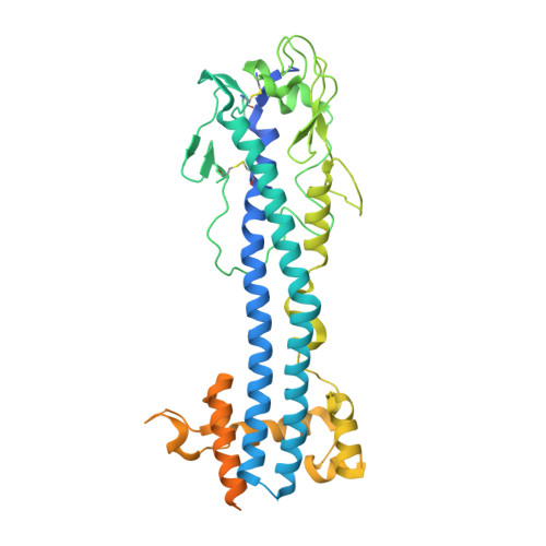
Variant Surface Glycoprotein 2 (VSG2, MiTat1.2, VSG221) Bound to Calcium


Experimental Data Snapshot

  • Method: X-RAY DIFFRACTION
  • Resolution: 1.74 Å
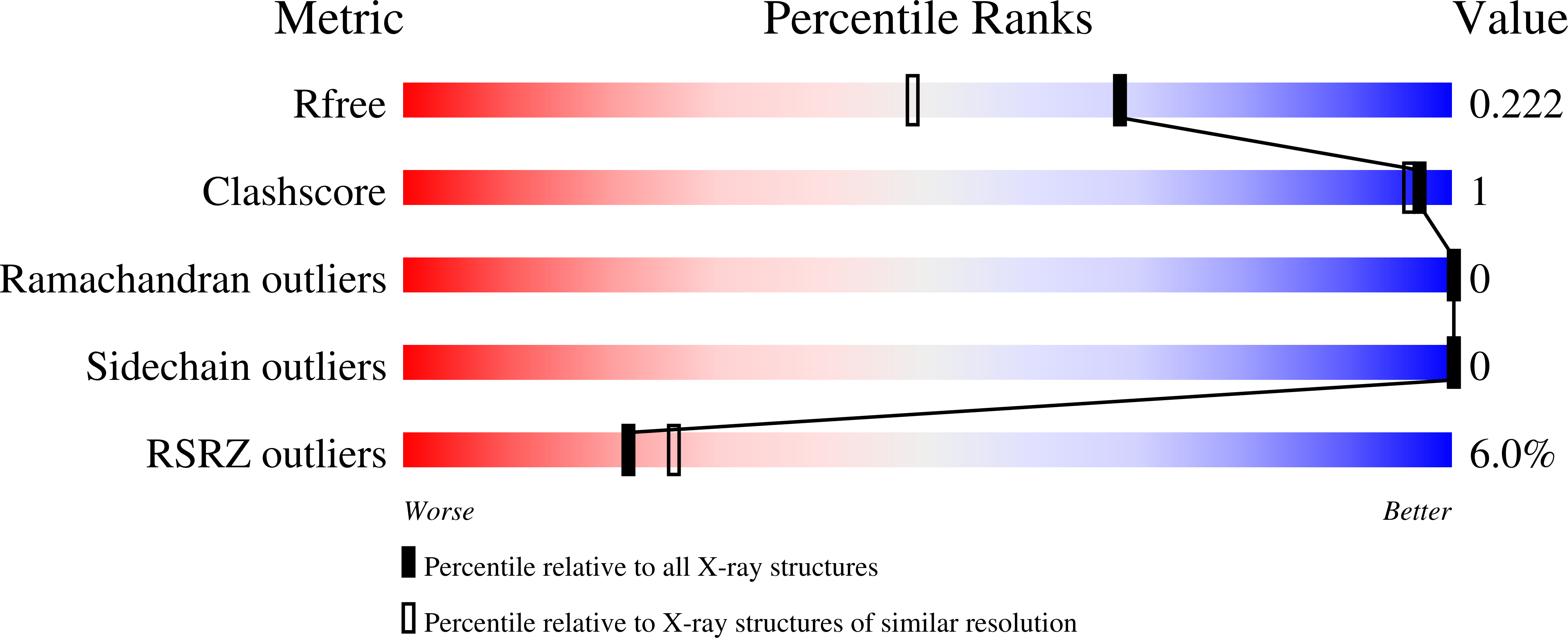
  • R-Value Free: 0.221 
  • R-Value Work: 0.189 
  • R-Value Observed: 0.191 

wwPDB Validation   3D Report Full Report


This is version 1.3 of the entry. See complete history

Re-refinement Note

This entry reflects an alternative modeling of the original data in: 1VSG


Literature

Immunodominant surface epitopes power immune evasion in the African trypanosome.

Gkeka, A.Aresta-Branco, F.Triller, G.Vlachou, E.P.van Straaten, M.Lilic, M.Olinares, P.D.B.Perez, K.Chait, B.T.Blatnik, R.Ruppert, T.Verdi, J.P.Stebbins, C.E.Papavasiliou, F.N.

(2023) Cell Rep 42: 112262-112262

  • DOI: https://doi.org/10.1016/j.celrep.2023.112262
  • Primary Citation of Related Structures:  
    7P56, 7P57, 7P59, 7P5A, 7P5B, 7P5D

  • PubMed Abstract: 

    The African trypanosome survives the immune response of its mammalian host by antigenic variation of its major surface antigen (the variant surface glycoprotein or VSG). Here we describe the antibody repertoires elicited by different VSGs. We show that the repertoires are highly restricted and are directed predominantly to distinct epitopes on the surface of the VSGs. They are also highly discriminatory; minor alterations within these exposed epitopes confer antigenically distinct properties to these VSGs and elicit different repertoires. We propose that the patterned and repetitive nature of the VSG coat focuses host immunity to a restricted set of immunodominant epitopes per VSG, eliciting a highly stereotyped response, minimizing cross-reactivity between different VSGs and facilitating prolonged immune evasion through epitope variation.


  • Organizational Affiliation

    Division of Immune Diversity, German Cancer Research Center, 69120 Heidelberg, Germany; Faculty of Biosciences, University of Heidelberg, 69120 Heidelberg, Germany; Panosome GmbH, 69123 Heidelberg, Germany.


Macromolecules
Find similar proteins by:  (by identity cutoff)  |  3D Structure
Entity ID: 1
MoleculeChains Sequence LengthOrganismDetailsImage
Variant surface glycoprotein MITAT 1.2
A, B
476Trypanosoma brucei bruceiMutation(s): 0 
UniProt
Find proteins for P26332 (Trypanosoma brucei brucei)
Explore P26332 
Go to UniProtKB:  P26332
Entity Groups  
Sequence Clusters30% Identity50% Identity70% Identity90% Identity95% Identity100% Identity
UniProt GroupP26332
Sequence Annotations
Expand
  • Reference Sequence
Oligosaccharides

Help

Entity ID: 2
MoleculeChains Length2D Diagram Glycosylation3D Interactions
beta-D-mannopyranose-(1-4)-2-acetamido-2-deoxy-beta-D-glucopyranose-(1-4)-2-acetamido-2-deoxy-beta-D-glucopyranose
C
3N-Glycosylation
Glycosylation Resources
GlyTouCan:  G15407YE
GlyCosmos:  G15407YE
GlyGen:  G15407YE
Entity ID: 3
MoleculeChains Length2D Diagram Glycosylation3D Interactions
alpha-D-mannopyranose-(1-6)-beta-D-mannopyranose-(1-4)-2-acetamido-2-deoxy-beta-D-glucopyranose-(1-4)-2-acetamido-2-deoxy-beta-D-glucopyranose
D
4N-Glycosylation
Glycosylation Resources
GlyTouCan:  G22573RC
GlyCosmos:  G22573RC
GlyGen:  G22573RC
Experimental Data & Validation

Experimental Data

  • Method: X-RAY DIFFRACTION
  • Resolution: 1.74 Å
  • R-Value Free: 0.221 
  • R-Value Work: 0.189 
  • R-Value Observed: 0.191 
  • Space Group: P 1 21 1
Unit Cell:
Length ( Å )Angle ( ˚ )
a = 42.915α = 90
b = 87.585β = 98.14
c = 88.121γ = 90
Software Package:
Software NamePurpose
PHENIXrefinement
PDB_EXTRACTdata extraction
XDSdata reduction
XDSdata scaling
PHENIXphasing

Structure Validation

View Full Validation Report


Entry History 

Deposition Data

Revision History  (Full details and data files)

  • Version 1.0: 2022-07-27
    Type: Initial release
  • Version 1.1: 2023-02-08
    Changes: Database references
  • Version 1.2: 2023-04-05
    Changes: Database references
  • Version 1.3: 2024-02-07
    Changes: Data collection, Refinement description