7BG1

Structure of anti-FLAG M2 Fab domain remodeled based on proteomic sequencing


Experimental Data Snapshot

  • Method: X-RAY DIFFRACTION
  • Resolution: 1.86 Å
  • R-Value Free: 0.255 
  • R-Value Work: 0.217 
  • R-Value Observed: 0.219 

wwPDB Validation   3D Report Full Report


This is version 1.1 of the entry. See complete history

Re-refinement Note

This entry reflects an alternative modeling of the original data in: 2G60


Literature

Mass Spectrometry-Based De Novo Sequencing of Monoclonal Antibodies Using Multiple Proteases and a Dual Fragmentation Scheme.

Peng, W.Pronker, M.F.Snijder, J.

(2021) J Proteome Res 20: 3559-3566

  • DOI: https://doi.org/10.1021/acs.jproteome.1c00169
  • Primary Citation of Related Structures:  
    7BG1

  • PubMed Abstract: 

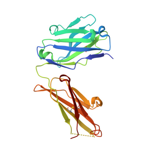
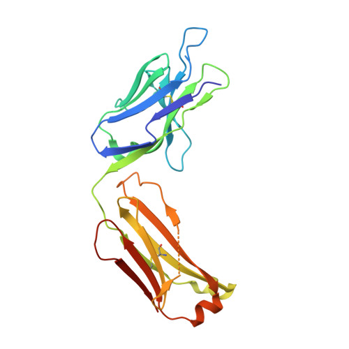
    Antibody sequence information is crucial to understanding the structural basis for antigen binding and enables the use of antibodies as therapeutics and research tools. Here, we demonstrate a method for direct de novo sequencing of monoclonal IgG from the purified antibody products. The method uses a panel of multiple complementary proteases to generate suitable peptides for de novo sequencing by liquid chromatography-tandem mass spectrometry (LC-MS/MS) in a bottom-up fashion. Furthermore, we apply a dual fragmentation scheme, using both stepped high-energy collision dissociation (stepped HCD) and electron-transfer high-energy collision dissociation (EThcD), on all peptide precursors. The method achieves full sequence coverage of the monoclonal antibody herceptin, with an accuracy of 99% in the variable regions. We applied the method to sequence the widely used anti-FLAG-M2 mouse monoclonal antibody, which we successfully validated by remodeling a high-resolution crystal structure of the Fab and demonstrating binding to a FLAG-tagged target protein in Western blot analysis. The method thus offers robust and reliable sequences of monoclonal antibodies.


  • Organizational Affiliation

    Biomolecular Mass Spectrometry and Proteomics, Bijvoet Center for Biomolecular Research and Utrecht Institute of Pharmaceutical Sciences, Utrecht University, Padualaan 8, 3584 CH Utrecht, The Netherlands.


Macromolecules
Find similar proteins by:  (by identity cutoff)  |  3D Structure
Entity ID: 1
MoleculeChains Sequence LengthOrganismDetailsImage
anti-FLAG M2 heavy chainA [auth H]222Mus musculusMutation(s): 0 
Gene Names: IGHV1-04/IGHJ2
Entity Groups  
Sequence Clusters30% Identity50% Identity70% Identity90% Identity95% Identity100% Identity
Sequence Annotations
Expand
  • Reference Sequence
Find similar proteins by:  (by identity cutoff)  |  3D Structure
Entity ID: 2
MoleculeChains Sequence LengthOrganismDetailsImage
anti-FLAG M2 light chainB [auth L]220Mus musculusMutation(s): 0 
Entity Groups  
Sequence Clusters30% Identity50% Identity70% Identity90% Identity95% Identity100% Identity
Sequence Annotations
Expand
  • Reference Sequence
Experimental Data & Validation

Experimental Data

  • Method: X-RAY DIFFRACTION
  • Resolution: 1.86 Å
  • R-Value Free: 0.255 
  • R-Value Work: 0.217 
  • R-Value Observed: 0.219 
  • Space Group: P 21 21 2
Unit Cell:
Length ( Å )Angle ( ˚ )
a = 87.36α = 90
b = 133.76β = 90
c = 41.48γ = 90
Software Package:
Software NamePurpose
Cootmodel building
PHENIXrefinement
DENZOdata reduction
SCALEPACKdata scaling
MOLREPphasing

Structure Validation

View Full Validation Report


Entry History & Funding Information

Deposition Data


Funding OrganizationLocationGrant Number
Netherlands Organisation for Scientific Research (NWO)Netherlands024.002.009

Revision History  (Full details and data files)

  • Version 1.0: 2021-07-21
    Type: Initial release
  • Version 1.1: 2024-01-31
    Changes: Data collection, Database references, Refinement description