Resolving the activation mechanism of the D99N antiterminator LicT protein.
Yang, Y., Gracy, J., Declerck, N., Demene, H.(2021) J Struct Biol 213: 107730-107730
- PubMed: 33781896
- DOI: https://doi.org/10.1016/j.jsb.2021.107730
- Primary Citation of Related Structures:
6TWR - PubMed Abstract:
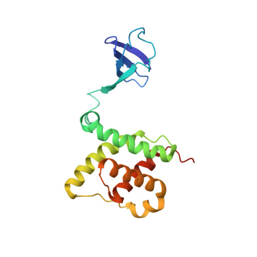
LicT is an antiterminator protein of the BglG family whose members are key players in the control of carbohydrate catabolism in bacteria. These antiterminators are generally composed of three modules, an N-terminal RNA-binding domain (CAT) followed by two homologous regulation modules (PRD1 and PRD2) that control the RNA binding activity of the effector domain via phosphorylation on conserved histidines. Although several structures of isolated domains of BglG-like proteins have been described, no structure containing CAT and at least one PRD simultaneously has yet been reported in an active state, precluding detailed understanding of signal transduction between modules. To fulfill this gap, we recently reported the complete NMR sequence assignment of a constitutively active mutant (D99N) CAT-PRD1*, which contains the effector domain and the first regulation domain of LicT. As a follow-up, we have determined and report here the 3D solution structure of this active, dimeric LicT construct (40 kDa). The structure reveals how the mutation constrains the PRD1 regulation domain into an active conformation which is transduced to CAT via a network of negatively charged residues belonging to PRD1 dimeric interface and to the linker region. In addition, our data support a model where BglG-type antitermination regulatory modules can only adopt a single conformation compatible with the active structure of the effector domain, regardless of whether activation is mediated by mutation on the first or second PRD. The linker between the effector and regulation modules appears to function as an adaptable hinge tuning the position of the functional modules.
Organizational Affiliation:
CBS, Univ Montpellier, CNRS, INSERM, Montpellier, France.