6T44

Ovine lactoglobulin complex with decanol


Experimental Data Snapshot

  • Method: X-RAY DIFFRACTION
  • Resolution: 2.00 Å
  • R-Value Free: 0.248 
  • R-Value Work: 0.196 
  • R-Value Observed: 0.199 

wwPDB Validation   3D Report Full Report


Ligand Structure Quality Assessment 


This is version 1.3 of the entry. See complete history


Literature

Conformational flexibility and ligand binding properties of ovine beta-lactoglobulin.

Loch, J.Bonarek, P.Lewinski, K.

(2019) Acta Biochim Pol 66: 577-584

  • DOI: https://doi.org/10.18388/abp.2019_2883
  • Primary Citation of Related Structures:  
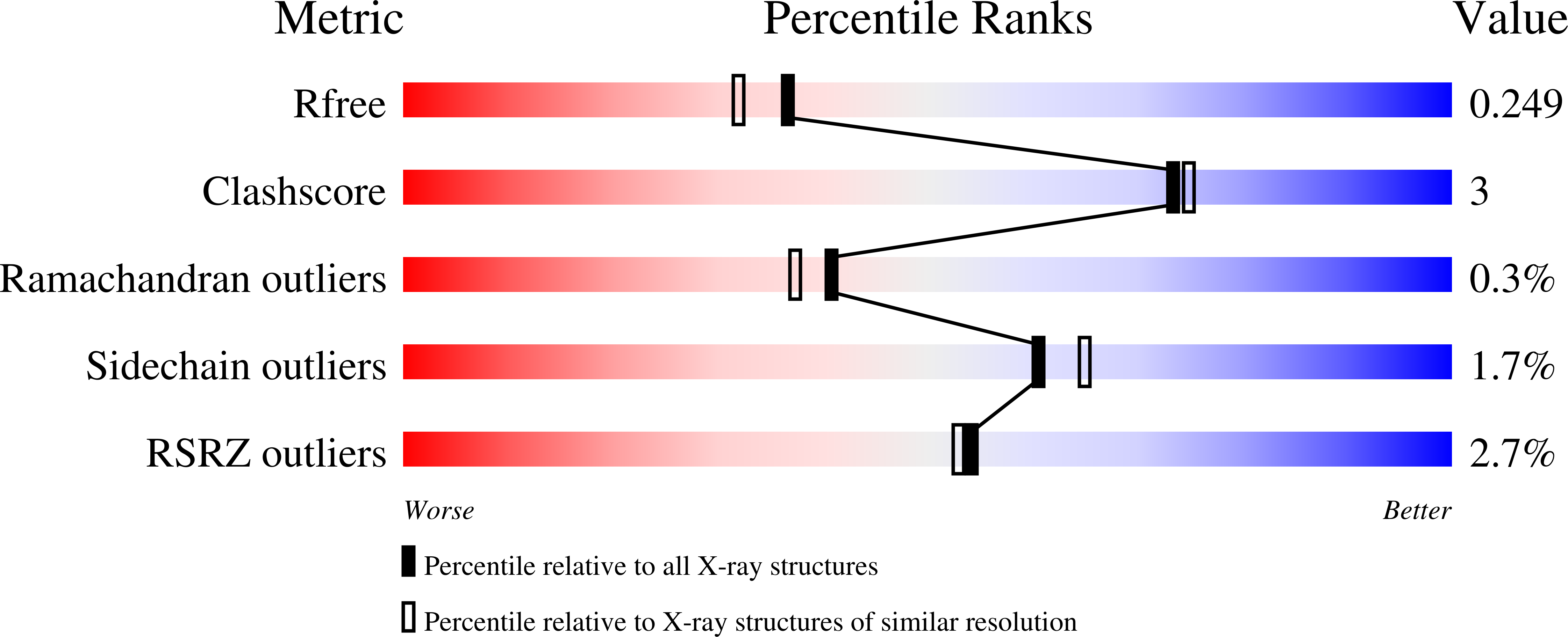
    6T42, 6T44

  • PubMed Abstract: 

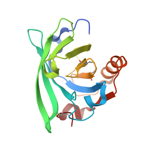
    Ovine β‑lactoglobulin was characterized by spectroscopic (CD), calorimetric (ITC) and X-ray structural studies. The structure of ovine β‑lactoglobulin complex with decanol showed that tight packing of molecules in the crystalline phase enforces a distortion of protein flexible loops resulting in the formation of an asymmetric dimer. The loops surrounding β-barrel in ovine lactoglobulin possessed the same conformational flexibility as observed previously in other lactoglobulins and the change of their conformation regulates the access to the binding pocket. The structure of asymmetric dimer revealed a new region in β-barrel where ligand polar group can be located. These findings indicated protein adaptability to ligand dimensions and inter- and intramolecular interactions in the crystalline phase. Calorimetric and crystallographic studies provided the experimental evidence that ovine lactoglobulin is able to bind aliphatic ligands. Thermodynamic parameters of sodium dodecyl sulfate binding determined by ITC at pH 7.5 had Ka, ΔH, TΔS and ΔG values similar to those observed for bovine and caprine protein indicating the same mechanism of ligand binding.


  • Organizational Affiliation

    Department of Crystal Chemistry and Crystal Physics, Faculty of Chemistry, Jagiellonian University, Kraków, Poland.


Macromolecules
Find similar proteins by:  (by identity cutoff)  |  3D Structure
Entity ID: 1
MoleculeChains Sequence LengthOrganismDetailsImage
Beta-lactoglobulin-1/BA [auth AAA],
B [auth BBB]
162Ovis ariesMutation(s): 1 
UniProt
Find proteins for P67976 (Ovis aries)
Explore P67976 
Go to UniProtKB:  P67976
Entity Groups  
Sequence Clusters30% Identity50% Identity70% Identity90% Identity95% Identity100% Identity
UniProt GroupP67976
Sequence Annotations
Expand
  • Reference Sequence
Experimental Data & Validation

Experimental Data

  • Method: X-RAY DIFFRACTION
  • Resolution: 2.00 Å
  • R-Value Free: 0.248 
  • R-Value Work: 0.196 
  • R-Value Observed: 0.199 
  • Space Group: P 62
Unit Cell:
Length ( Å )Angle ( ˚ )
a = 108.34α = 90
b = 108.34β = 90
c = 55.856γ = 120
Software Package:
Software NamePurpose
REFMACrefinement
CrysalisProdata reduction
Aimlessdata scaling
PHASERphasing

Structure Validation

View Full Validation Report


Ligand Structure Quality Assessment 


Entry History & Funding Information

Deposition Data


Funding OrganizationLocationGrant Number
Polish National Science CentrePoland2012/05/B/ST5/00278

Revision History  (Full details and data files)

  • Version 1.0: 2019-10-23
    Type: Initial release
  • Version 1.1: 2020-01-08
    Changes: Database references
  • Version 1.2: 2020-01-15
    Changes: Database references
  • Version 1.3: 2024-01-24
    Changes: Data collection, Database references, Derived calculations, Refinement description