6SJ5

Crystal structure of the uL14-RsfS complex from Staphylococcus aureus


Experimental Data Snapshot

  • Method: X-RAY DIFFRACTION
  • Resolution: 2.27 Å
  • R-Value Free: 0.264 
  • R-Value Work: 0.229 
  • R-Value Observed: 0.230 

wwPDB Validation   3D Report Full Report


This is version 1.1 of the entry. See complete history


Literature

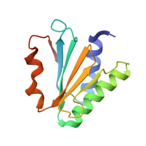
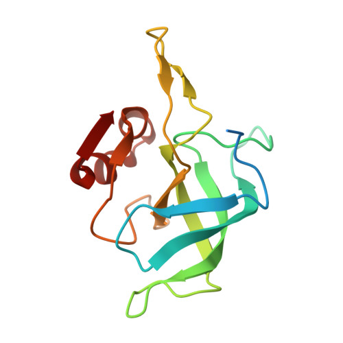
Mechanism of ribosome shutdown by RsfS in Staphylococcus aureus revealed by integrative structural biology approach.

Khusainov, I.Fatkhullin, B.Pellegrino, S.Bikmullin, A.Liu, W.T.Gabdulkhakov, A.Shebel, A.A.Golubev, A.Zeyer, D.Trachtmann, N.Sprenger, G.A.Validov, S.Usachev, K.Yusupova, G.Yusupov, M.

(2020) Nat Commun 11: 1656-1656

  • DOI: https://doi.org/10.1038/s41467-020-15517-0
  • Primary Citation of Related Structures:  
    6SJ5, 6SJ6

  • PubMed Abstract: 

    For the sake of energy preservation, bacteria, upon transition to stationary phase, tone down their protein synthesis. This process is favored by the reversible binding of small stress-induced proteins to the ribosome to prevent unnecessary translation. One example is the conserved bacterial ribosome silencing factor (RsfS) that binds to uL14 protein onto the large ribosomal subunit and prevents its association with the small subunit. Here we describe the binding mode of Staphylococcus aureus RsfS to the large ribosomal subunit and present a 3.2 Å resolution cryo-EM reconstruction of the 50S-RsfS complex together with the crystal structure of uL14-RsfS complex solved at 2.3 Å resolution. The understanding of the detailed landscape of RsfS-uL14 interactions within the ribosome shed light on the mechanism of ribosome shutdown in the human pathogen S. aureus and might deliver a novel target for pharmacological drug development and treatment of bacterial infections.


  • Organizational Affiliation

    Laboratory of Structural Biology, Institute of Fundamental Medicine and Biology, Kazan Federal University, Kremlyovskaya Street 18, Kazan, 420008, Russia. iskander.khusainov@biophys.mpg.de.


Macromolecules
Find similar proteins by:  (by identity cutoff)  |  3D Structure
Entity ID: 1
MoleculeChains Sequence LengthOrganismDetailsImage
Ribosomal silencing factor RsfS
A, B
117Staphylococcus aureusMutation(s): 0 
Gene Names: rsfSE5491_08675FAF14_04505FAF30_12305FAF31_00945FAF32_10770QU38_08975
UniProt
Find proteins for W8TVV3 (Staphylococcus aureus)
Explore W8TVV3 
Go to UniProtKB:  W8TVV3
Entity Groups  
Sequence Clusters30% Identity50% Identity70% Identity90% Identity95% Identity100% Identity
UniProt GroupW8TVV3
Sequence Annotations
Expand
  • Reference Sequence
Find similar proteins by:  (by identity cutoff)  |  3D Structure
Entity ID: 2
MoleculeChains Sequence LengthOrganismDetailsImage
50S ribosomal protein L14
C, D
142Staphylococcus aureus subsp. aureus NCTC 8325Mutation(s): 0 
Gene Names: rplNSAOUHSC_02502
UniProt
Find proteins for Q2FW16 (Staphylococcus aureus (strain NCTC 8325 / PS 47))
Explore Q2FW16 
Go to UniProtKB:  Q2FW16
Entity Groups  
Sequence Clusters30% Identity50% Identity70% Identity90% Identity95% Identity100% Identity
UniProt GroupQ2FW16
Sequence Annotations
Expand
  • Reference Sequence
Experimental Data & Validation

Experimental Data

  • Method: X-RAY DIFFRACTION
  • Resolution: 2.27 Å
  • R-Value Free: 0.264 
  • R-Value Work: 0.229 
  • R-Value Observed: 0.230 
  • Space Group: P 21 21 21
Unit Cell:
Length ( Å )Angle ( ˚ )
a = 37.107α = 90
b = 117.601β = 90
c = 121.217γ = 90
Software Package:
Software NamePurpose
PHENIXrefinement
XDSdata reduction
XSCALEdata scaling
PHASERphasing

Structure Validation

View Full Validation Report


Entry History 

Deposition Data

Revision History  (Full details and data files)

  • Version 1.0: 2020-04-15
    Type: Initial release
  • Version 1.1: 2024-01-24
    Changes: Data collection, Database references, Refinement description