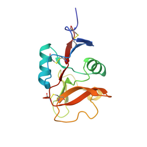
6LFJ

Crystal structure of mouse DCAR2 CRD domain complex with IPM2


Experimental Data Snapshot

  • Method: X-RAY DIFFRACTION
  • Resolution: 1.84 Å
  • R-Value Free: 0.198 
  • R-Value Work: 0.177 
  • R-Value Observed: 0.178 

wwPDB Validation   3D Report Full Report


Ligand Structure Quality Assessment 


This is version 1.3 of the entry. See complete history


Literature

Structural insight into the recognition of pathogen-derived phosphoglycolipids by C-type lectin receptor DCAR.

Omahdi, Z.Horikawa, Y.Nagae, M.Toyonaga, K.Imamura, A.Takato, K.Teramoto, T.Ishida, H.Kakuta, Y.Yamasaki, S.

(2020) J Biol Chem 295: 5807-5817

  • DOI: https://doi.org/10.1074/jbc.RA120.012491
  • Primary Citation of Related Structures:  
    6KZR, 6LFJ, 6LKR

  • PubMed Abstract: 

    The C-type lectin receptors (CLRs) form a family of pattern recognition receptors that recognize numerous pathogens, such as bacteria and fungi, and trigger innate immune responses. The extracellular carbohydrate-recognition domain (CRD) of CLRs forms a globular structure that can coordinate a Ca 2+ ion, allowing receptor interactions with sugar-containing ligands. Although well-conserved, the CRD fold can also display differences that directly affect the specificity of the receptors for their ligands. Here, we report crystal structures at 1.8-2.3 Å resolutions of the CRD of murine dendritic cell-immunoactivating receptor (DCAR, or Clec4b1 ), the CLR that binds phosphoglycolipids such as acylated phosphatidyl- myo -inositol mannosides (AcPIMs) of mycobacteria. Using mutagenesis analysis, we identified critical residues, Ala 136 and Gln 198 , on the surface surrounding the ligand-binding site of DCAR, as well as an atypical Ca 2+ -binding motif (Glu-Pro-Ser/EPS 168-170 ). By chemically synthesizing a water-soluble ligand analog, inositol-monophosphate dimannose (IPM2), we confirmed the direct interaction of DCAR with the polar moiety of AcPIMs by biolayer interferometry and co-crystallization approaches. We also observed a hydrophobic groove extending from the ligand-binding site that is in a suitable position to interact with the lipid portion of whole AcPIMs. These results suggest that the hydroxyl group-binding ability and hydrophobic groove of DCAR mediate its specific binding to pathogen-derived phosphoglycolipids such as mycobacterial AcPIMs.


  • Organizational Affiliation

    Department of Molecular Immunology, Research Institute for Microbial Diseases, Osaka University, Suita 565-0871, Japan; Laboratory of Molecular Immunology, Immunology Frontier Research Center (IFReC), Osaka University, Suita 565-0871, Japan; Division of Molecular Immunology, Medical Institute of Bioregulation, Kyushu University, Fukuoka 812-8582, Japan.


Macromolecules
Find similar proteins by:  (by identity cutoff)  |  3D Structure
Entity ID: 1
MoleculeChains Sequence LengthOrganismDetailsImage
C-type lectin domain family 4, member b1
A, B
136Mus musculusMutation(s): 0 
Gene Names: Clec4b1Clec4bDcar
UniProt
Find proteins for Q9D8Q7 (Mus musculus)
Explore Q9D8Q7 
Go to UniProtKB:  Q9D8Q7
Entity Groups  
Sequence Clusters30% Identity50% Identity70% Identity90% Identity95% Identity100% Identity
UniProt GroupQ9D8Q7
Sequence Annotations
Expand
  • Reference Sequence
Small Molecules
Ligands 4 Unique
IDChains Name / Formula / InChI Key2D Diagram3D Interactions
B3P
Query on B3P

Download Ideal Coordinates CCD File 
D [auth A]2-[3-(2-HYDROXY-1,1-DIHYDROXYMETHYL-ETHYLAMINO)-PROPYLAMINO]-2-HYDROXYMETHYL-PROPANE-1,3-DIOL
C11 H26 N2 O6
HHKZCCWKTZRCCL-UHFFFAOYSA-N
IPD
Query on IPD

Download Ideal Coordinates CCD File 
F [auth A],
I [auth B]
D-MYO-INOSITOL-1-PHOSPHATE
C6 H11 O9 P
INAPMGSXUVUWAF-UOTPTPDRSA-L
MAN
Query on MAN

Download Ideal Coordinates CCD File 
E [auth A],
G [auth A],
J [auth B]
alpha-D-mannopyranose
C6 H12 O6
WQZGKKKJIJFFOK-PQMKYFCFSA-N
CA
Query on CA

Download Ideal Coordinates CCD File 
C [auth A],
H [auth B]
CALCIUM ION
Ca
BHPQYMZQTOCNFJ-UHFFFAOYSA-N
Experimental Data & Validation

Experimental Data

  • Method: X-RAY DIFFRACTION
  • Resolution: 1.84 Å
  • R-Value Free: 0.198 
  • R-Value Work: 0.177 
  • R-Value Observed: 0.178 
  • Space Group: P 21 21 21
Unit Cell:
Length ( Å )Angle ( ˚ )
a = 65.689α = 90
b = 72.564β = 90
c = 100.469γ = 90
Software Package:
Software NamePurpose
PHENIXrefinement
XDSdata reduction
XDSdata scaling
MOLREPphasing

Structure Validation

View Full Validation Report


Ligand Structure Quality Assessment 


Entry History 

Deposition Data

Revision History  (Full details and data files)

  • Version 1.0: 2020-03-25
    Type: Initial release
  • Version 1.1: 2020-05-06
    Changes: Database references
  • Version 1.2: 2020-07-29
    Type: Remediation
    Reason: Carbohydrate remediation
    Changes: Data collection, Derived calculations, Structure summary
  • Version 1.3: 2023-11-22
    Changes: Data collection, Database references, Derived calculations, Refinement description, Structure summary