6GEW

OphA Y63F-sinefungin complex


Experimental Data Snapshot

  • Method: X-RAY DIFFRACTION
  • Resolution: 2.10 Å
  • R-Value Free: 0.258 
  • R-Value Work: 0.235 
  • R-Value Observed: 0.236 

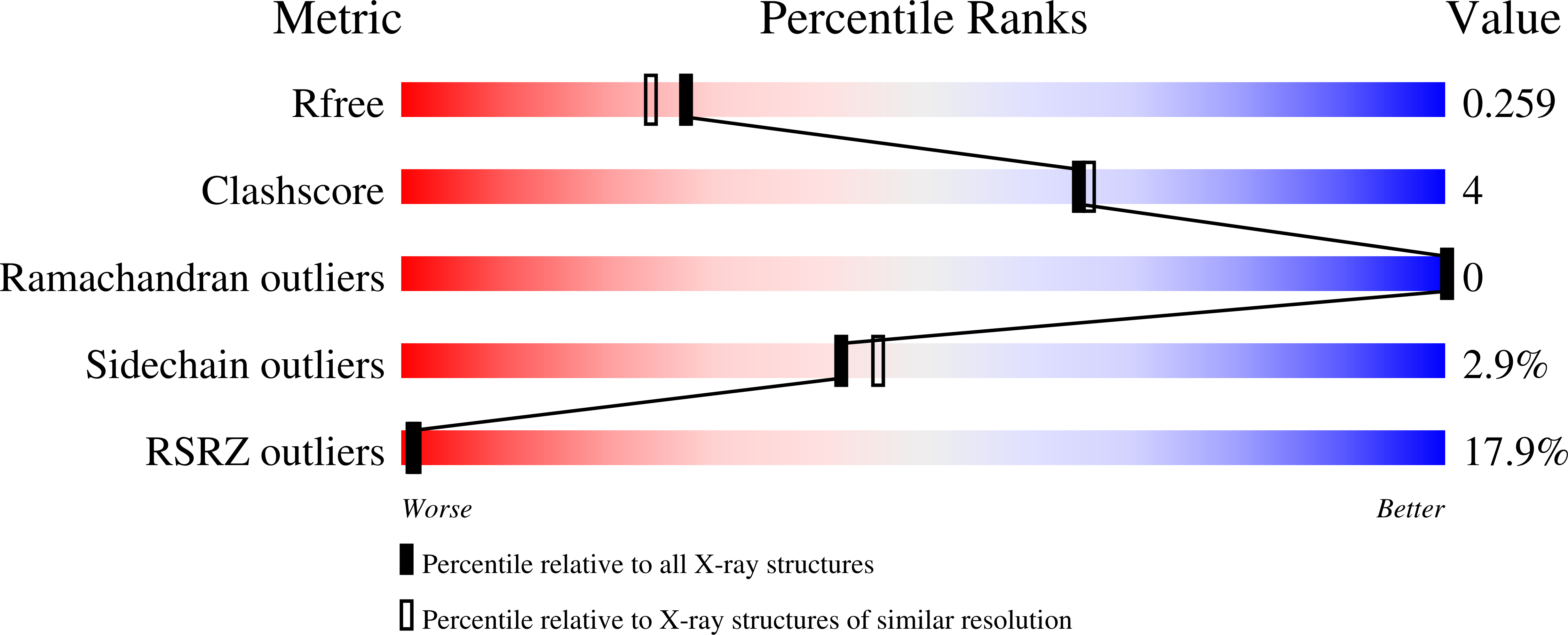
wwPDB Validation   3D Report Full Report


Ligand Structure Quality Assessment 


This is version 1.1 of the entry. See complete history


Literature

A molecular mechanism for the enzymatic methylation of nitrogen atoms within peptide bonds.

Song, H.van der Velden, N.S.Shiran, S.L.Bleiziffer, P.Zach, C.Sieber, R.Imani, A.S.Krausbeck, F.Aebi, M.Freeman, M.F.Riniker, S.Kunzler, M.Naismith, J.H.

(2018) Sci Adv 4: eaat2720-eaat2720

  • DOI: https://doi.org/10.1126/sciadv.aat2720
  • Primary Citation of Related Structures:  
    5N0N, 5N0O, 5N0P, 5N0Q, 5N0R, 5N0S, 5N0T, 5N0U, 5N0V, 5N0W, 5N0X, 5N4I, 5OUF, 6GEW

  • PubMed Abstract: 

    The peptide bond, the defining feature of proteins, governs peptide chemistry by abolishing nucleophilicity of the nitrogen. This and the planarity of the peptide bond arise from the delocalization of the lone pair of electrons on the nitrogen atom into the adjacent carbonyl. While chemical methylation of an amide bond uses a strong base to generate the imidate, OphA, the precursor protein of the fungal peptide macrocycle omphalotin A, self-hypermethylates amides at pH 7 using S -adenosyl methionine (SAM) as cofactor. The structure of OphA reveals a complex catenane-like arrangement in which the peptide substrate is clamped with its amide nitrogen aligned for nucleophilic attack on the methyl group of SAM. Biochemical data and computational modeling suggest a base-catalyzed reaction with the protein stabilizing the reaction intermediate. Backbone N-methylation of peptides enhances their protease resistance and membrane permeability, a property that holds promise for applications to medicinal chemistry.


  • Organizational Affiliation

    Biomedical Sciences Research Complex, North Haugh, University of St. Andrews, Fife KY16 9ST, UK.


Macromolecules
Find similar proteins by:  (by identity cutoff)  |  3D Structure
Entity ID: 1
MoleculeChains Sequence LengthOrganismDetailsImage
OphA410Omphalotus oleariusMutation(s): 0 
UniProt
Find proteins for A0A2R2JFI5 (Omphalotus olearius)
Explore A0A2R2JFI5 
Go to UniProtKB:  A0A2R2JFI5
Entity Groups  
Sequence Clusters30% Identity50% Identity70% Identity90% Identity95% Identity100% Identity
UniProt GroupA0A2R2JFI5
Sequence Annotations
Expand
  • Reference Sequence
Small Molecules
Ligands 2 Unique
IDChains Name / Formula / InChI Key2D Diagram3D Interactions
SAH
Query on SAH

Download Ideal Coordinates CCD File 
B [auth A]S-ADENOSYL-L-HOMOCYSTEINE
C14 H20 N6 O5 S
ZJUKTBDSGOFHSH-WFMPWKQPSA-N
SFG
Query on SFG

Download Ideal Coordinates CCD File 
C [auth A]SINEFUNGIN
C15 H23 N7 O5
LMXOHSDXUQEUSF-YECHIGJVSA-N
Modified Residues  1 Unique
IDChains TypeFormula2D DiagramParent
MVA
Query on MVA
A
L-PEPTIDE LINKINGC6 H13 N O2VAL
Experimental Data & Validation

Experimental Data

  • Method: X-RAY DIFFRACTION
  • Resolution: 2.10 Å
  • R-Value Free: 0.258 
  • R-Value Work: 0.235 
  • R-Value Observed: 0.236 
  • Space Group: P 32 2 1
Unit Cell:
Length ( Å )Angle ( ˚ )
a = 89.784α = 90
b = 89.784β = 90
c = 156.902γ = 120
Software Package:
Software NamePurpose
REFMACrefinement
xia2data reduction
xia2data scaling
PHASERphasing

Structure Validation

View Full Validation Report


Ligand Structure Quality Assessment 


Entry History 

Deposition Data

Revision History  (Full details and data files)

  • Version 1.0: 2018-09-19
    Type: Initial release
  • Version 1.1: 2024-01-17
    Changes: Data collection, Database references, Refinement description