6EYT

Crystal structure of the Salmonella effector SseK3 in complex with UDP-GlcNAc and Manganese


Experimental Data Snapshot

  • Method: X-RAY DIFFRACTION
  • Resolution: 2.21 Å
  • R-Value Free: 0.241 
  • R-Value Work: 0.196 
  • R-Value Observed: 0.199 

wwPDB Validation   3D Report Full Report


Ligand Structure Quality Assessment 


This is version 2.0 of the entry. See complete history


Literature

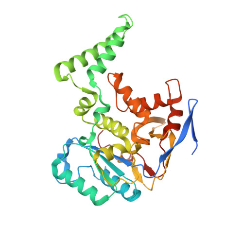
Structural basis for the glycosyltransferase activity of theSalmonellaeffector SseK3.

Esposito, D.Gunster, R.A.Martino, L.El Omari, K.Wagner, A.Thurston, T.L.M.Rittinger, K.

(2018) J Biol Chem 293: 5064-5078

  • DOI: https://doi.org/10.1074/jbc.RA118.001796
  • Primary Citation of Related Structures:  
    6EYR, 6EYT

  • PubMed Abstract: 

    The Salmonella -secreted effector SseK3 translocates into host cells, targeting innate immune responses, including NF-κB activation. SseK3 is a glycosyltransferase that transfers an N -acetylglucosamine (GlcNAc) moiety onto the guanidino group of a target arginine, modulating host cell function. However, a lack of structural information has precluded elucidation of the molecular mechanisms in arginine and GlcNAc selection. We report here the crystal structure of SseK3 in its apo form and in complex with hydrolyzed UDP-GlcNAc. SseK3 possesses the typical glycosyltransferase type-A (GT-A)-family fold and the metal-coordinating D X D motif essential for ligand binding and enzymatic activity. Several conserved residues were essential for arginine GlcNAcylation and SseK3-mediated inhibition of NF-κB activation. Isothermal titration calorimetry revealed SseK3's preference for manganese coordination. The pattern of interactions in the substrate-bound SseK3 structure explained the selection of the primary ligand. Structural rearrangement of the C-terminal residues upon ligand binding was crucial for SseK3's catalytic activity, and NMR analysis indicated that SseK3 has limited UDP-GlcNAc hydrolysis activity. The release of free N -acetyl α-d-glucosamine, and the presence of the same molecule in the SseK3 active site, classified it as a retaining glycosyltransferase. A glutamate residue in the active site suggested a double-inversion mechanism for the arginine N -glycosylation reaction. Homology models of SseK1, SseK2, and the Escherichia coli orthologue NleB1 reveal differences in the surface electrostatic charge distribution, possibly accounting for their diverse activities. This first structure of a retaining GT-A arginine N -glycosyltransferase provides an important step toward a better understanding of this enzyme class and their roles as bacterial effectors.


  • Organizational Affiliation

    From the Molecular Structure of Cell Signalling Laboratory, Francis Crick Institute, 1 Midland Road, London NW1 1AT, United Kingdom.


Macromolecules
Find similar proteins by:  (by identity cutoff)  |  3D Structure
Entity ID: 1
MoleculeChains Sequence LengthOrganismDetailsImage
Type III secretion system effector protein
A, B
327Salmonella enterica subsp. enterica serovar Typhimurium str. SL1344Mutation(s): 0 
Gene Names: sseK3SL1344_1928
UniProt
Find proteins for A0A0H3NMP8 (Salmonella typhimurium (strain SL1344))
Explore A0A0H3NMP8 
Go to UniProtKB:  A0A0H3NMP8
Entity Groups  
Sequence Clusters30% Identity50% Identity70% Identity90% Identity95% Identity100% Identity
UniProt GroupA0A0H3NMP8
Sequence Annotations
Expand
  • Reference Sequence
Small Molecules
Ligands 4 Unique
IDChains Name / Formula / InChI Key2D Diagram3D Interactions
UDP
Query on UDP

Download Ideal Coordinates CCD File 
F [auth A],
K [auth B]
URIDINE-5'-DIPHOSPHATE
C9 H14 N2 O12 P2
XCCTYIAWTASOJW-XVFCMESISA-N
NDG
Query on NDG

Download Ideal Coordinates CCD File 
G [auth A],
L [auth B]
2-acetamido-2-deoxy-alpha-D-glucopyranose
C8 H15 N O6
OVRNDRQMDRJTHS-PVFLNQBWSA-N
IOD
Query on IOD

Download Ideal Coordinates CCD File 
C [auth A],
D [auth A],
H [auth B],
I [auth B]
IODIDE ION
I
XMBWDFGMSWQBCA-UHFFFAOYSA-M
MN
Query on MN

Download Ideal Coordinates CCD File 
E [auth A],
J [auth B]
MANGANESE (II) ION
Mn
WAEMQWOKJMHJLA-UHFFFAOYSA-N
Experimental Data & Validation

Experimental Data

  • Method: X-RAY DIFFRACTION
  • Resolution: 2.21 Å
  • R-Value Free: 0.241 
  • R-Value Work: 0.196 
  • R-Value Observed: 0.199 
  • Space Group: P 21 21 21
Unit Cell:
Length ( Å )Angle ( ˚ )
a = 73.97α = 90
b = 85.94β = 90
c = 96.03γ = 90
Software Package:
Software NamePurpose
PHENIXrefinement
XDSdata reduction
XDSdata scaling
MOLREPphasing

Structure Validation

View Full Validation Report


Ligand Structure Quality Assessment 


Entry History & Funding Information

Deposition Data


Funding OrganizationLocationGrant Number
The Francis Crick InstituteUnited KingdomFC001142

Revision History  (Full details and data files)

  • Version 1.0: 2018-02-28
    Type: Initial release
  • Version 1.1: 2018-04-18
    Changes: Data collection, Database references
  • Version 2.0: 2020-07-29
    Type: Remediation
    Reason: Carbohydrate remediation
    Changes: Advisory, Atomic model, Data collection, Derived calculations, Structure summary