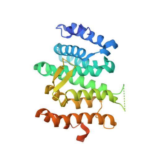
6EMG

Crystal structure of Rrp1 from Chaetomium thermophilum in space group P6322


Experimental Data Snapshot

  • Method: X-RAY DIFFRACTION
  • Resolution: 2.24 Å
  • R-Value Free: 0.207 
  • R-Value Work: 0.187 
  • R-Value Observed: 0.188 

wwPDB Validation   3D Report Full Report


This is version 1.2 of the entry. See complete history


Literature

Visualizing the Assembly Pathway of Nucleolar Pre-60S Ribosomes.

Kater, L.Thoms, M.Barrio-Garcia, C.Cheng, J.Ismail, S.Ahmed, Y.L.Bange, G.Kressler, D.Berninghausen, O.Sinning, I.Hurt, E.Beckmann, R.

(2017) Cell 171: 1599-1610.e14

  • DOI: https://doi.org/10.1016/j.cell.2017.11.039
  • Primary Citation of Related Structures:  
    6ELZ, 6EM1, 6EM3, 6EM4, 6EM5, 6EMF, 6EMG, 6EN7

  • PubMed Abstract: 

    Eukaryotic 60S ribosomal subunits are comprised of three rRNAs and ∼50 ribosomal proteins. The initial steps of their formation take place in the nucleolus, but, owing to a lack of structural information, this process is poorly understood. Using cryo-EM, we solved structures of early 60S biogenesis intermediates at 3.3 Å to 4.5 Å resolution, thereby providing insights into their sequential folding and assembly pathway. Besides revealing distinct immature rRNA conformations, we map 25 assembly factors in six different assembly states. Notably, the Nsa1-Rrp1-Rpf1-Mak16 module stabilizes the solvent side of the 60S subunit, and the Erb1-Ytm1-Nop7 complex organizes and connects through Erb1's meandering N-terminal extension, eight assembly factors, three ribosomal proteins, and three 25S rRNA domains. Our structural snapshots reveal the order of integration and compaction of the six major 60S domains within early nucleolar 60S particles developing stepwise from the solvent side around the exit tunnel to the central protuberance.


  • Organizational Affiliation

    Gene Center Munich and Center of Integrated Protein Science-Munich (CiPS-M), Department of Biochemistry, Feodor-Lynen-Str. 25, University of Munich, 81377 Munich, Germany.


Macromolecules
Find similar proteins by:  (by identity cutoff)  |  3D Structure
Entity ID: 1
MoleculeChains Sequence LengthOrganismDetailsImage
G0S4M2
A, B
290Thermochaetoides thermophila DSM 1495Mutation(s): 0 
Gene Names: CTHT_0031510
UniProt
Find proteins for G0S4M2 (Chaetomium thermophilum (strain DSM 1495 / CBS 144.50 / IMI 039719))
Explore G0S4M2 
Go to UniProtKB:  G0S4M2
Entity Groups  
Sequence Clusters30% Identity50% Identity70% Identity90% Identity95% Identity100% Identity
UniProt GroupG0S4M2
Sequence Annotations
Expand
  • Reference Sequence
Small Molecules
Ligands 3 Unique
IDChains Name / Formula / InChI Key2D Diagram3D Interactions
EPE
Query on EPE

Download Ideal Coordinates CCD File 
L [auth A]4-(2-HYDROXYETHYL)-1-PIPERAZINE ETHANESULFONIC ACID
C8 H18 N2 O4 S
JKMHFZQWWAIEOD-UHFFFAOYSA-N
PO4
Query on PO4

Download Ideal Coordinates CCD File 
C [auth A],
D [auth A],
M [auth B],
N [auth B]
PHOSPHATE ION
O4 P
NBIIXXVUZAFLBC-UHFFFAOYSA-K
EDO
Query on EDO

Download Ideal Coordinates CCD File 
E [auth A]
F [auth A]
G [auth A]
H [auth A]
I [auth A]
E [auth A],
F [auth A],
G [auth A],
H [auth A],
I [auth A],
J [auth A],
K [auth A]
1,2-ETHANEDIOL
C2 H6 O2
LYCAIKOWRPUZTN-UHFFFAOYSA-N
Experimental Data & Validation

Experimental Data

  • Method: X-RAY DIFFRACTION
  • Resolution: 2.24 Å
  • R-Value Free: 0.207 
  • R-Value Work: 0.187 
  • R-Value Observed: 0.188 
  • Space Group: P 63 2 2
Unit Cell:
Length ( Å )Angle ( ˚ )
a = 214.259α = 90
b = 214.259β = 90
c = 124.785γ = 120
Software Package:
Software NamePurpose
XDSdata reduction
Aimlessdata scaling
SHELXDEphasing
PHENIXrefinement
PDB_EXTRACTdata extraction

Structure Validation

View Full Validation Report


Entry History & Funding Information

Deposition Data


Funding OrganizationLocationGrant Number
German Research FoundationGermany--

Revision History  (Full details and data files)

  • Version 1.0: 2017-12-27
    Type: Initial release
  • Version 1.1: 2018-01-31
    Changes: Author supporting evidence
  • Version 1.2: 2019-10-16
    Changes: Data collection