6COJ

Crystal structure of Rhodococcus jostii RHA1 IpdAB E105A COCHEA-COA complex


Experimental Data Snapshot

  • Method: X-RAY DIFFRACTION
  • Resolution: 1.40 Å
  • R-Value Free: 0.173 
  • R-Value Work: 0.152 
  • R-Value Observed: 0.153 

wwPDB Validation   3D Report Full Report


Ligand Structure Quality Assessment 


This is version 1.4 of the entry. See complete history


Literature

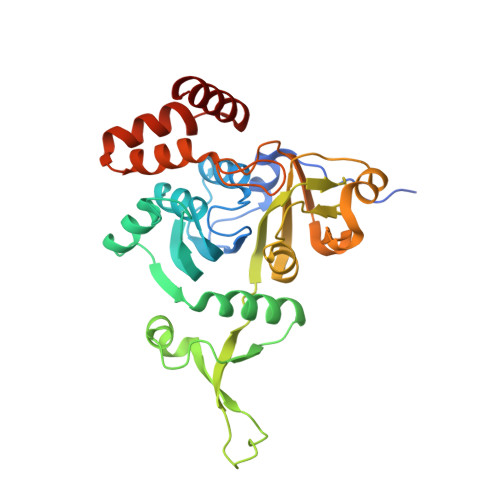
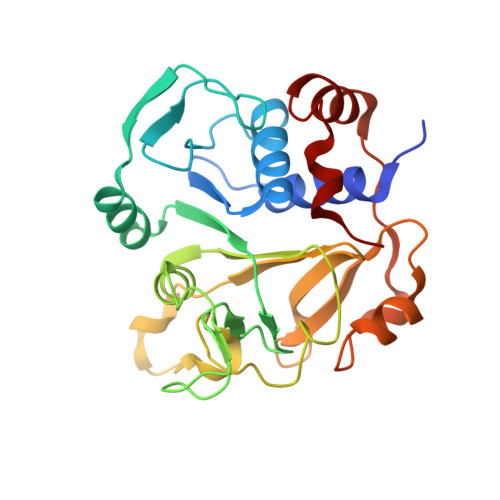
IpdAB, a virulence factor inMycobacterium tuberculosis, is a cholesterol ring-cleaving hydrolase.

Crowe, A.M.Workman, S.D.Watanabe, N.Worrall, L.J.Strynadka, N.C.J.Eltis, L.D.

(2018) Proc Natl Acad Sci U S A 115: E3378-E3387

  • DOI: https://doi.org/10.1073/pnas.1717015115
  • Primary Citation of Related Structures:  
    6CO6, 6CO9, 6COJ, 6CON

  • PubMed Abstract: 

    Mycobacterium tuberculosis ( Mtb ) grows on host-derived cholesterol during infection. IpdAB, found in all steroid-degrading bacteria and a determinant of pathogenicity, has been implicated in the hydrolysis of the last steroid ring. Phylogenetic analyses revealed that IpdAB orthologs form a clade of CoA transferases (CoTs). In a coupled assay with a thiolase, IpdAB transformed the cholesterol catabolite ( R )-2-(2-carboxyethyl)-3-methyl-6-oxocyclohex-1-ene-1-carboxyl-CoA (COCHEA-CoA) and CoASH to 4-methyl-5-oxo-octanedioyl-CoA (MOODA-CoA) and acetyl-CoA with high specificity ( k cat / K m = 5.8 ± 0.8 × 10 4 M -1 ⋅s -1 ). The structure of MOODA-CoA was consistent with IpdAB hydrolyzing COCHEA-CoA to a β-keto-thioester, a thiolase substrate. Contrary to characterized CoTs, IpdAB exhibited no activity toward small CoA thioesters. Further, IpdAB lacks the catalytic glutamate residue that is conserved in the β-subunit of characterized CoTs and a glutamyl-CoA intermediate was not trapped during turnover. By contrast, Glu105 A , conserved in the α-subunit of IpdAB, was essential for catalysis. A crystal structure of the IpdAB·COCHEA-CoA complex, solved to 1.4 Å, revealed that Glu105 A is positioned to act as a catalytic base. Upon titration with COCHEA-CoA, the E105A A variant accumulated a yellow-colored species (λ max = 310 nm; K d = 0.4 ± 0.2 μM) typical of β-keto enolates. In the presence of D 2 O, IpdAB catalyzed the deuteration of COCHEA-CoA adjacent to the hydroxylation site at rates consistent with k cat Based on these data and additional IpdAB variants, we propose a retro-Claisen condensation-like mechanism for the IpdAB-mediated hydrolysis of COCHEA-CoA. This study expands the range of known reactions catalyzed by the CoT superfamily and provides mechanistic insight into an important determinant of Mtb pathogenesis.


  • Organizational Affiliation

    Department of Biochemistry and Molecular Biology, Life Sciences Institute, The University of British Columbia, Vancouver, BC, Canada, V6T 1Z3.


Macromolecules
Find similar proteins by:  (by identity cutoff)  |  3D Structure
Entity ID: 1
MoleculeChains Sequence LengthOrganismDetailsImage
Probable CoA-transferase alpha subunit308Rhodococcus jostii RHA1Mutation(s): 1 
Gene Names: RHA1_ro04651
UniProt
Find proteins for Q0S7P9 (Rhodococcus jostii (strain RHA1))
Explore Q0S7P9 
Go to UniProtKB:  Q0S7P9
Entity Groups  
Sequence Clusters30% Identity50% Identity70% Identity90% Identity95% Identity100% Identity
UniProt GroupQ0S7P9
Sequence Annotations
Expand
  • Reference Sequence
Find similar proteins by:  (by identity cutoff)  |  3D Structure
Entity ID: 2
MoleculeChains Sequence LengthOrganismDetailsImage
Probable CoA-transferase beta subunit253Rhodococcus jostii RHA1Mutation(s): 0 
Gene Names: RHA1_ro04650
UniProt
Find proteins for Q0S7Q0 (Rhodococcus jostii (strain RHA1))
Explore Q0S7Q0 
Go to UniProtKB:  Q0S7Q0
Entity Groups  
Sequence Clusters30% Identity50% Identity70% Identity90% Identity95% Identity100% Identity
UniProt GroupQ0S7Q0
Sequence Annotations
Expand
  • Reference Sequence
Small Molecules
Ligands 2 Unique
IDChains Name / Formula / InChI Key2D Diagram3D Interactions
F8G (Subject of Investigation/LOI)
Query on F8G

Download Ideal Coordinates CCD File 
E [auth B]S-{(3R,5R,9R)-1-[(2R,3S,4R,5R)-5-(6-amino-9H-purin-9-yl)-4-hydroxy-3-(phosphonooxy)tetrahydrofuran-2-yl]-3,5,9-trihydroxy-8,8-dimethyl-3,5-dioxido-10,14-dioxo-2,4,6-trioxa-11,15-diaza-3lambda~5~,5lambda~5~-diphosphaheptadecan-17-yl} (5R,10R)-7-hydroxy-10-methyl-2-oxo-1-oxaspiro[4.5]dec-6-ene-6-carbothioate (non-preferred name)
C32 H48 N7 O20 P3 S
COMHLEMSBYWTLA-LJKJHTHQSA-N
SO4
Query on SO4

Download Ideal Coordinates CCD File 
C [auth A],
D [auth A],
F [auth B]
SULFATE ION
O4 S
QAOWNCQODCNURD-UHFFFAOYSA-L
Experimental Data & Validation

Experimental Data

  • Method: X-RAY DIFFRACTION
  • Resolution: 1.40 Å
  • R-Value Free: 0.173 
  • R-Value Work: 0.152 
  • R-Value Observed: 0.153 
  • Space Group: P 43 21 2
Unit Cell:
Length ( Å )Angle ( ˚ )
a = 69.172α = 90
b = 69.172β = 90
c = 241.873γ = 90
Software Package:
Software NamePurpose
PHENIXrefinement
Aimlessdata scaling
PDB_EXTRACTdata extraction
XDSdata reduction
PHASERphasing

Structure Validation

View Full Validation Report


Ligand Structure Quality Assessment 


Entry History 

Deposition Data

Revision History  (Full details and data files)

  • Version 1.0: 2018-03-28
    Type: Initial release
  • Version 1.1: 2018-04-11
    Changes: Data collection, Database references
  • Version 1.2: 2018-04-25
    Changes: Data collection, Database references
  • Version 1.3: 2018-06-13
    Changes: Data collection, Structure summary
  • Version 1.4: 2023-10-04
    Changes: Data collection, Database references, Refinement description