6CN9

Crystal structure of the Kinase domain of WNK1


Experimental Data Snapshot

  • Method: X-RAY DIFFRACTION
  • Resolution: 1.80 Å
  • R-Value Free: 0.211 
  • R-Value Work: 0.161 
  • R-Value Observed: 0.164 

wwPDB Validation   3D Report Full Report


This is version 1.1 of the entry. See complete history

Re-refinement Note

This entry reflects an alternative modeling of the original data in: 3FPQ


Literature

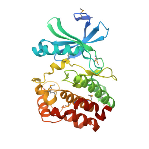
Crystal structure of the kinase domain of WNK1, a kinase that causes a hereditary form of hypertension.

Min, X.Lee, B.H.Cobb, M.H.Goldsmith, E.J.

(2004) Structure 12: 1303-1311

  • DOI: https://doi.org/10.1016/j.str.2004.04.014
  • Primary Citation of Related Structures:  
    6CN9

  • PubMed Abstract: 

    WNK kinases comprise a small group of unique serine/threonine protein kinases that have been genetically linked to pseudohypoaldosteronism type II, an autosomal dominant form of hypertension. Here we present the structure of the kinase domain of WNK1 at 1.8 A resolution, solved in a low activity conformation. A lysine residue (Lys-233) is found in the active site emanating from strand beta2 rather than strand beta3 as in other protein kinases. The activation loop adopts a unique well-folded inactive conformation. The conformations of the P+1 specificity pocket, the placement of the conserved active site threonine (Thr-386), and the exterior placement of helix C, contribute to the low activity state. By homology modeling, we identified two hydrophobic residues in the substrate-binding groove that contribute to substrate specificity. The structure of the WNK1 catalytic domain, with its unique active site, may help in the design of therapeutic reagents for the treatment of hypertension.


  • Organizational Affiliation

    Department of Biochemistry, University of Texas Southwestern Medical Center at Dallas, 5323 Harry Hines Boulevard, Dallas, TX 75390, USA.


Macromolecules
Find similar proteins by:  (by identity cutoff)  |  3D Structure
Entity ID: 1
MoleculeChains Sequence LengthOrganismDetailsImage
Serine/threonine-protein kinase WNK1
A, B
290Rattus norvegicusMutation(s): 0 
Gene Names: Wnk1Hsn2Prkwnk1
EC: 2.7.11.1
UniProt
Find proteins for Q9JIH7 (Rattus norvegicus)
Explore Q9JIH7 
Go to UniProtKB:  Q9JIH7
Entity Groups  
Sequence Clusters30% Identity50% Identity70% Identity90% Identity95% Identity100% Identity
UniProt GroupQ9JIH7
Sequence Annotations
Expand
  • Reference Sequence
Small Molecules
Modified Residues  1 Unique
IDChains TypeFormula2D DiagramParent
MSE
Query on MSE
A, B
L-PEPTIDE LINKINGC5 H11 N O2 SeMET
Experimental Data & Validation

Experimental Data

  • Method: X-RAY DIFFRACTION
  • Resolution: 1.80 Å
  • R-Value Free: 0.211 
  • R-Value Work: 0.161 
  • R-Value Observed: 0.164 
  • Space Group: P 1
Unit Cell:
Length ( Å )Angle ( ˚ )
a = 38.308α = 91.32
b = 57.767β = 89.99
c = 65.657γ = 90.88
Software Package:
Software NamePurpose
REFMACrefinement
HKL-2000data reduction
HKL-2000data scaling
SOLVEphasing

Structure Validation

View Full Validation Report


Entry History & Funding Information

Deposition Data


Funding OrganizationLocationGrant Number
National Institutes of Health/National Institute of Diabetes and Digestive and Kidney Disease (NIH/NIDDK)United States--

Revision History  (Full details and data files)

  • Version 1.0: 2019-03-27
    Type: Initial release
  • Version 1.1: 2019-12-25
    Changes: Author supporting evidence