6CEN

Crystal Structure of WHSC1L1 in Complex with Inhibitor PEP21


Experimental Data Snapshot

  • Method: X-RAY DIFFRACTION
  • Resolution: 1.61 Å
  • R-Value Free: 0.215 
  • R-Value Work: 0.179 
  • R-Value Observed: 0.181 

wwPDB Validation   3D Report Full Report


Ligand Structure Quality Assessment 


This is version 1.1 of the entry. See complete history


Literature

Identification of a peptide inhibitor for the histone methyltransferase WHSC1.

Morrison, M.J.Boriack-Sjodin, P.A.Swinger, K.K.Wigle, T.J.Sadalge, D.Kuntz, K.W.Scott, M.P.Janzen, W.P.Chesworth, R.Duncan, K.W.Harvey, D.M.Lampe, J.W.Mitchell, L.H.Copeland, R.A.

(2018) PLoS One 13: e0197082-e0197082

  • DOI: https://doi.org/10.1371/journal.pone.0197082
  • Primary Citation of Related Structures:  
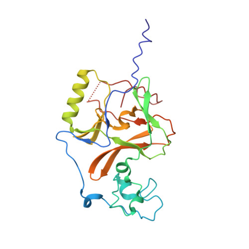
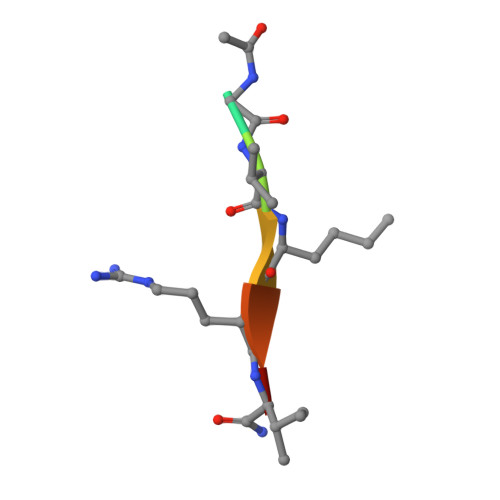
    6CEN

  • PubMed Abstract: 

    WHSC1 is a histone methyltransferase that is responsible for mono- and dimethylation of lysine 36 on histone H3 and has been implicated as a driver in a variety of hematological and solid tumors. Currently, there is a complete lack of validated chemical matter for this important drug discovery target. Herein we report on the first fully validated WHSC1 inhibitor, PTD2, a norleucine-containing peptide derived from the histone H4 sequence. This peptide exhibits micromolar affinity towards WHSC1 in biochemical and biophysical assays. Furthermore, a crystal structure was solved with the peptide in complex with SAM and the SET domain of WHSC1L1. This inhibitor is an important first step in creating potent, selective WHSC1 tool compounds for the purposes of understanding the complex biology in relation to human disease.


  • Organizational Affiliation

    Epizyme Inc., Cambridge, Massachusetts, United States of America.


Macromolecules
Find similar proteins by:  (by identity cutoff)  |  3D Structure
Entity ID: 1
MoleculeChains Sequence LengthOrganismDetailsImage
Histone-lysine N-methyltransferase NSD3228Homo sapiensMutation(s): 0 
Gene Names: NSD3WHSC1L1DC28
EC: 2.1.1.43
UniProt & NIH Common Fund Data Resources
Find proteins for Q9BZ95 (Homo sapiens)
Explore Q9BZ95 
Go to UniProtKB:  Q9BZ95
PHAROS:  Q9BZ95
GTEx:  ENSG00000147548 
Entity Groups  
Sequence Clusters30% Identity50% Identity70% Identity90% Identity95% Identity100% Identity
UniProt GroupQ9BZ95
Sequence Annotations
Expand
  • Reference Sequence

Find similar proteins by:  Sequence   |   3D Structure  

Entity ID: 2
MoleculeChains Sequence LengthOrganismDetailsImage
ACE-GLY-VAL-NLE-ARG-ILE-NH2B [auth D]7Homo sapiensMutation(s): 0 
Sequence Annotations
Expand
  • Reference Sequence
Experimental Data & Validation

Experimental Data

  • Method: X-RAY DIFFRACTION
  • Resolution: 1.61 Å
  • R-Value Free: 0.215 
  • R-Value Work: 0.179 
  • R-Value Observed: 0.181 
  • Space Group: P 1 21 1
Unit Cell:
Length ( Å )Angle ( ˚ )
a = 42.87α = 90
b = 62.66β = 107.92
c = 48.6γ = 90
Software Package:
Software NamePurpose
REFMACrefinement
PDB_EXTRACTdata extraction

Structure Validation

View Full Validation Report


Ligand Structure Quality Assessment 


Entry History 

Deposition Data

Revision History  (Full details and data files)

  • Version 1.0: 2018-05-09
    Type: Initial release
  • Version 1.1: 2018-05-23
    Changes: Data collection, Database references