6B0N

Crystal structure of the cleavage-independent prefusion HIV Env glycoprotein trimer of the clade A BG505 isolate (NFL construct) in complex with Fabs PGT122 and PGV19 at 3.39 A


Experimental Data Snapshot

  • Method: X-RAY DIFFRACTION
  • Resolution: 3.40 Å
  • R-Value Free: 0.346 
  • R-Value Work: 0.335 
  • R-Value Observed: 0.336 

wwPDB Validation   3D Report Full Report


Ligand Structure Quality Assessment 


This is version 2.1 of the entry. See complete history


Literature

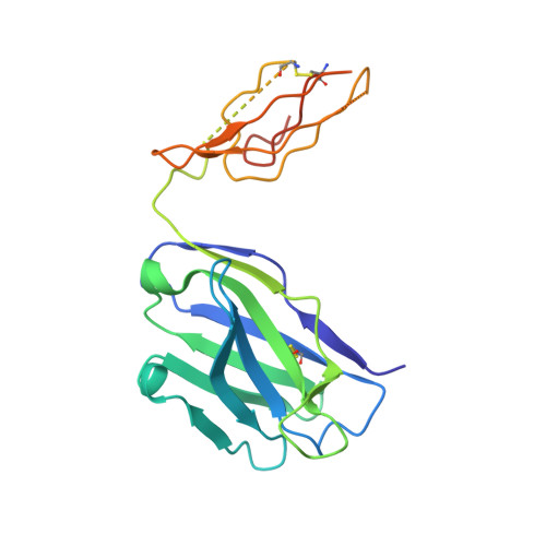
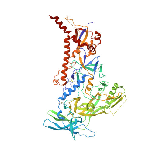
Structure of a cleavage-independent HIV Env recapitulates the glycoprotein architecture of the native cleaved trimer.

Sarkar, A.Bale, S.Behrens, A.J.Kumar, S.Sharma, S.K.de Val, N.Pallesen, J.Irimia, A.Diwanji, D.C.Stanfield, R.L.Ward, A.B.Crispin, M.Wyatt, R.T.Wilson, I.A.

(2018) Nat Commun 9: 1956-1956

  • DOI: https://doi.org/10.1038/s41467-018-04272-y
  • Primary Citation of Related Structures:  
    6AVN, 6B0N

  • PubMed Abstract: 

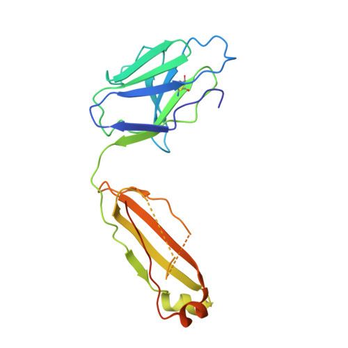
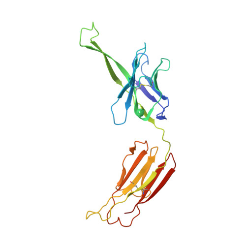
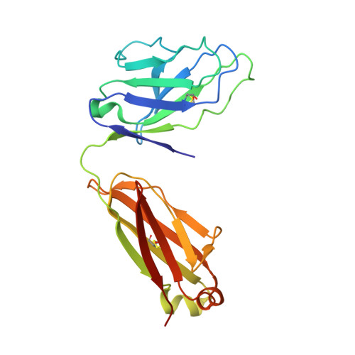
    Furin cleavage of the HIV envelope glycoprotein is an essential step for cell entry that enables formation of well-folded, native-like glycosylated trimers, releases constraints on the fusion peptide, and limits enzymatic processing of the N-glycan shield. Here, we show that a cleavage-independent, stabilized, soluble Env trimer mimic (BG505 NFL.664) exhibits a "closed-form", native-like, prefusion conformation akin to furin-cleaved Env trimers. The crystal structure of BG505 NFL.664 at 3.39 Å resolution with two potent bNAbs also identifies the full epitopes of PGV19 and PGT122 that target the receptor binding site and N332 supersite, respectively. Quantitative site-specific analysis of the glycan shield reveals that native-like glycan processing is maintained despite furin-independent maturation in the secretory pathway. Thus, cleavage-independent NFL Env trimers exhibit quaternary protein and carbohydrate structures similar to the native viral spike that further validate their potential as vaccine immunogen candidates.


  • Organizational Affiliation

    IAVI Neutralizing Antibody Center, The Scripps Research Institute, La Jolla, CA, 92037, USA.


Macromolecules
Find similar proteins by:  (by identity cutoff)  |  3D Structure
Entity ID: 1
MoleculeChains Sequence LengthOrganismDetailsImage
PGV19 Fab heavy chainA [auth D]223Homo sapiensMutation(s): 0 
Entity Groups  
Sequence Clusters30% Identity50% Identity70% Identity90% Identity95% Identity100% Identity
Sequence Annotations
Expand
  • Reference Sequence
Find similar proteins by:  (by identity cutoff)  |  3D Structure
Entity ID: 2
MoleculeChains Sequence LengthOrganismDetailsImage
PGV19 light chainB [auth E]209Homo sapiensMutation(s): 0 
Entity Groups  
Sequence Clusters30% Identity50% Identity70% Identity90% Identity95% Identity100% Identity
Sequence Annotations
Expand
  • Reference Sequence
Find similar proteins by:  (by identity cutoff)  |  3D Structure
Entity ID: 3
MoleculeChains Sequence LengthOrganismDetailsImage
Envelope glycoprotein gp140C [auth G]638Human immunodeficiency virus 1Mutation(s): 2 
Gene Names: env
UniProt
Find proteins for Q2N0S6 (Human immunodeficiency virus 1)
Explore Q2N0S6 
Go to UniProtKB:  Q2N0S6
Entity Groups  
Sequence Clusters30% Identity50% Identity70% Identity90% Identity95% Identity100% Identity
UniProt GroupQ2N0S6
Sequence Annotations
Expand
  • Reference Sequence
Find similar proteins by:  (by identity cutoff)  |  3D Structure
Entity ID: 4
MoleculeChains Sequence LengthOrganismDetailsImage
PGT122 Fab heavy chainD [auth H]235Homo sapiensMutation(s): 0 
Entity Groups  
Sequence Clusters30% Identity50% Identity70% Identity90% Identity95% Identity100% Identity
Sequence Annotations
Expand
  • Reference Sequence
Find similar proteins by:  (by identity cutoff)  |  3D Structure
Entity ID: 5
MoleculeChains Sequence LengthOrganismDetailsImage
PGT122 light chainE [auth L]213Homo sapiensMutation(s): 0 
Entity Groups  
Sequence Clusters30% Identity50% Identity70% Identity90% Identity95% Identity100% Identity
Sequence Annotations
Expand
  • Reference Sequence
Oligosaccharides

Help

Entity ID: 6
MoleculeChains Length2D Diagram Glycosylation3D Interactions
alpha-D-mannopyranose-(1-2)-alpha-D-mannopyranose-(1-3)-[alpha-D-mannopyranose-(1-3)-alpha-D-mannopyranose-(1-6)]beta-D-mannopyranose-(1-4)-2-acetamido-2-deoxy-beta-D-glucopyranose-(1-4)-2-acetamido-2-deoxy-beta-D-glucopyranoseF [auth A]7N-Glycosylation
Glycosylation Resources
GlyTouCan:  G07617FP
GlyCosmos:  G07617FP
GlyGen:  G07617FP
Entity ID: 7
MoleculeChains Length2D Diagram Glycosylation3D Interactions
beta-D-mannopyranose-(1-4)-2-acetamido-2-deoxy-beta-D-glucopyranose-(1-4)-2-acetamido-2-deoxy-beta-D-glucopyranoseG [auth B],
H [auth C]
3N-Glycosylation
Glycosylation Resources
GlyTouCan:  G15407YE
GlyCosmos:  G15407YE
GlyGen:  G15407YE
Entity ID: 8
MoleculeChains Length2D Diagram Glycosylation3D Interactions
alpha-D-mannopyranose-(1-2)-alpha-D-mannopyranose-(1-2)-alpha-D-mannopyranose-(1-3)-[alpha-D-mannopyranose-(1-6)-alpha-D-mannopyranose-(1-6)]beta-D-mannopyranose-(1-4)-2-acetamido-2-deoxy-beta-D-glucopyranose-(1-4)-2-acetamido-2-deoxy-beta-D-glucopyranoseI [auth F]8N-Glycosylation
Glycosylation Resources
GlyTouCan:  G81980VO
GlyCosmos:  G81980VO
GlyGen:  G81980VO
Entity ID: 9
MoleculeChains Length2D Diagram Glycosylation3D Interactions
alpha-D-mannopyranose-(1-3)-[alpha-D-mannopyranose-(1-6)]beta-D-mannopyranose-(1-4)-2-acetamido-2-deoxy-beta-D-glucopyranose-(1-4)-2-acetamido-2-deoxy-beta-D-glucopyranoseJ [auth I],
O
5N-Glycosylation
Glycosylation Resources
GlyTouCan:  G22768VO
GlyCosmos:  G22768VO
GlyGen:  G22768VO
Entity ID: 10
MoleculeChains Length2D Diagram Glycosylation3D Interactions
2-acetamido-2-deoxy-beta-D-glucopyranose-(1-4)-2-acetamido-2-deoxy-beta-D-glucopyranoseK [auth J],
P,
R
2N-Glycosylation
Glycosylation Resources
GlyTouCan:  G42666HT
GlyCosmos:  G42666HT
GlyGen:  G42666HT
Entity ID: 11
MoleculeChains Length2D Diagram Glycosylation3D Interactions
alpha-D-mannopyranose-(1-6)-beta-D-mannopyranose-(1-4)-2-acetamido-2-deoxy-beta-D-glucopyranose-(1-4)-2-acetamido-2-deoxy-beta-D-glucopyranoseL [auth K]4N-Glycosylation
Glycosylation Resources
GlyTouCan:  G22573RC
GlyCosmos:  G22573RC
GlyGen:  G22573RC
Entity ID: 12
MoleculeChains Length2D Diagram Glycosylation3D Interactions
alpha-D-mannopyranose-(1-2)-alpha-D-mannopyranose-(1-3)-[alpha-D-mannopyranose-(1-2)-alpha-D-mannopyranose-(1-6)]alpha-D-mannopyranose-(1-6)-beta-D-mannopyranose-(1-4)-2-acetamido-2-deoxy-beta-D-glucopyranose-(1-4)-2-acetamido-2-deoxy-beta-D-glucopyranose
M
8N-Glycosylation
Glycosylation Resources
GlyTouCan:  G48018RF
GlyCosmos:  G48018RF
GlyGen:  G48018RF
Entity ID: 13
MoleculeChains Length2D Diagram Glycosylation3D Interactions
alpha-D-mannopyranose-(1-2)-alpha-D-mannopyranose-(1-2)-alpha-D-mannopyranose
N
3N/A
Glycosylation Resources
GlyTouCan:  G25559RS
GlyCosmos:  G25559RS
GlyGen:  G25559RS
Entity ID: 14
MoleculeChains Length2D Diagram Glycosylation3D Interactions
alpha-D-mannopyranose-(1-3)-alpha-D-mannopyranose-(1-6)-[alpha-D-mannopyranose-(1-3)]beta-D-mannopyranose
Q
4N/A
Glycosylation Resources
GlyTouCan:  G65169RI
GlyCosmos:  G65169RI
GlyGen:  G65169RI
Entity ID: 15
MoleculeChains Length2D Diagram Glycosylation3D Interactions
alpha-D-mannopyranose-(1-2)-alpha-D-mannopyranose-(1-3)-[alpha-D-mannopyranose-(1-3)-[alpha-D-mannopyranose-(1-6)]alpha-D-mannopyranose-(1-6)]beta-D-mannopyranose-(1-4)-2-acetamido-2-deoxy-beta-D-glucopyranose-(1-4)-2-acetamido-2-deoxy-beta-D-glucopyranose
S
8N-Glycosylation
Glycosylation Resources
GlyTouCan:  G80966KZ
GlyCosmos:  G80966KZ
GlyGen:  G80966KZ
Small Molecules
Ligands 2 Unique
IDChains Name / Formula / InChI Key2D Diagram3D Interactions
NAG
Query on NAG

Download Ideal Coordinates CCD File 
AA [auth G]
BA [auth G]
CA [auth G]
DA [auth G]
EA [auth G]
AA [auth G],
BA [auth G],
CA [auth G],
DA [auth G],
EA [auth G],
FA [auth G],
T [auth G],
U [auth G],
W [auth G],
X [auth G],
Y [auth G],
Z [auth G]
2-acetamido-2-deoxy-beta-D-glucopyranose
C8 H15 N O6
OVRNDRQMDRJTHS-FMDGEEDCSA-N
MAN
Query on MAN

Download Ideal Coordinates CCD File 
GA [auth G],
V [auth G]
alpha-D-mannopyranose
C6 H12 O6
WQZGKKKJIJFFOK-PQMKYFCFSA-N
Experimental Data & Validation

Experimental Data

  • Method: X-RAY DIFFRACTION
  • Resolution: 3.40 Å
  • R-Value Free: 0.346 
  • R-Value Work: 0.335 
  • R-Value Observed: 0.336 
  • Space Group: P 63
Unit Cell:
Length ( Å )Angle ( ˚ )
a = 161.247α = 90
b = 161.247β = 90
c = 245.651γ = 120
Software Package:
Software NamePurpose
PHENIXrefinement
HKL-2000data reduction
HKL-2000data scaling
PHASERphasing

Structure Validation

View Full Validation Report


Ligand Structure Quality Assessment 


Entry History 

Deposition Data

Revision History  (Full details and data files)

  • Version 1.0: 2018-05-30
    Type: Initial release
  • Version 2.0: 2020-07-29
    Type: Remediation
    Reason: Carbohydrate remediation
    Changes: Advisory, Atomic model, Data collection, Derived calculations, Structure summary
  • Version 2.1: 2023-10-04
    Changes: Data collection, Database references, Refinement description, Structure summary