5ZDR

Crystal structure of cyanide-insensitive alternative oxidase from Trypanosoma brucei with ascofuranone derivative


Experimental Data Snapshot

  • Method: X-RAY DIFFRACTION
  • Resolution: 2.59 Å
  • R-Value Free: 0.257 
  • R-Value Work: 0.206 
  • R-Value Observed: 0.209 

wwPDB Validation   3D Report Full Report


Ligand Structure Quality Assessment 


This is version 1.3 of the entry. See complete history

Re-refinement Note

This entry reflects an alternative modeling of the original data in: 3VVA


Literature

Insights into the ubiquinol/dioxygen binding and proton relay pathways of the alternative oxidase.

Shiba, T.Inaoka, D.K.Takahashi, G.Tsuge, C.Kido, Y.Young, L.Ueda, S.Balogun, E.O.Nara, T.Honma, T.Tanaka, A.Inoue, M.Saimoto, H.Harada, S.Moore, A.L.Kita, K.

(2019) Biochim Biophys Acta Bioenerg 1860: 375-382

  • DOI: https://doi.org/10.1016/j.bbabio.2019.03.008
  • Primary Citation of Related Structures:  
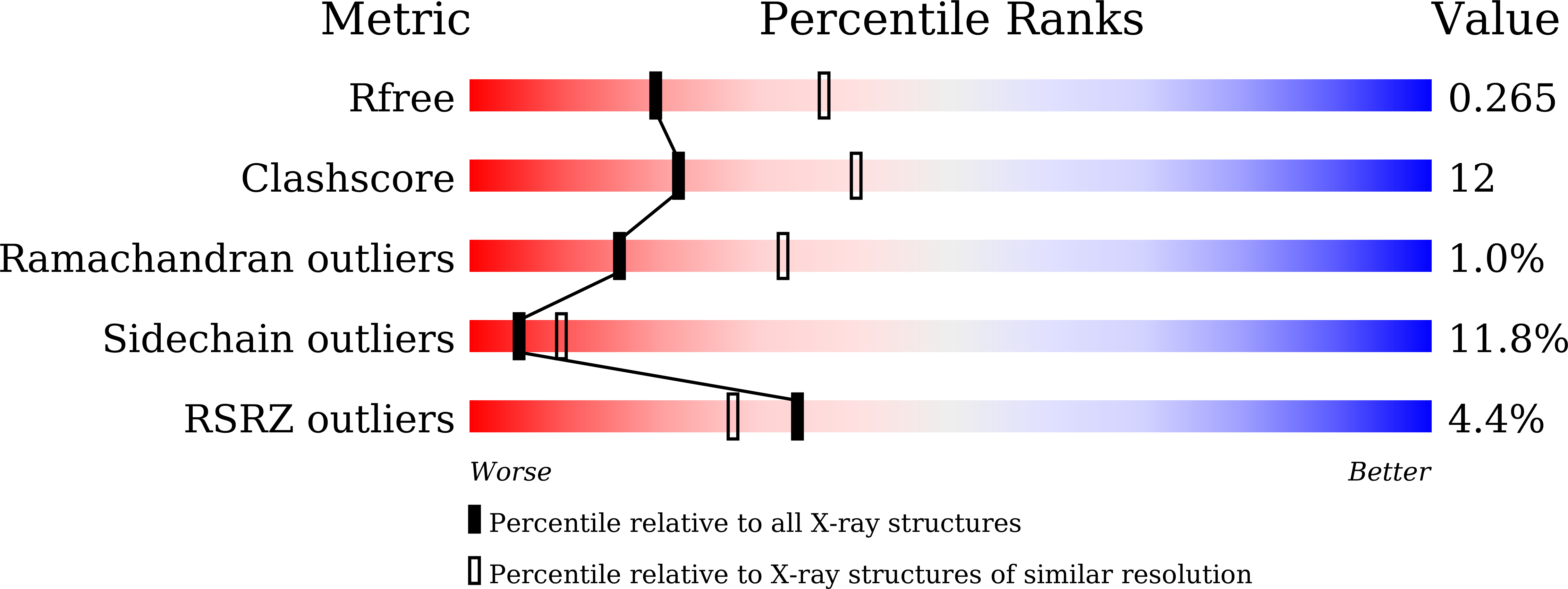
    5ZDP, 5ZDQ, 5ZDR

  • PubMed Abstract: 

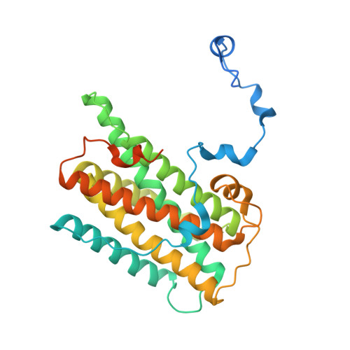
    The alternative oxidase (AOX) is a monotopic diiron carboxylate protein which catalyzes the four-electron reduction of dioxygen to water by ubiquinol. Although we have recently determined the crystal structure of Trypanosoma brucei AOX (TAO) in the presence and absence of ascofuranone (AF) derivatives (which are potent mixed type inhibitors) the mechanism by which ubiquinol and dioxygen binds to TAO remain inconclusive. In this article, ferulenol was identified as the first competitive inhibitor of AOX which has been used to probe the binding of ubiquinol. Surface plasmon resonance reveals that AF is a quasi-irreversible inhibitor of TAO whilst ferulenol binding is completely reversible. The structure of the TAO-ferulenol complex, determined at 2.7 Å, provided insights into ubiquinol binding and has also identified a potential dioxygen molecule bound in a side-on conformation to the diiron center for the first time.


  • Organizational Affiliation

    Department of Applied Biology, Graduate School of Science and Technology, Kyoto Institute of Technology, Sakyo-ku, Matsugasaki, Kyoto 606-8585, Japan. Electronic address: tshiba@kit.ac.jp.


Macromolecules
Find similar proteins by:  (by identity cutoff)  |  3D Structure
Entity ID: 1
MoleculeChains Sequence LengthOrganismDetailsImage
Alternative oxidase, mitochondrial
A, B, C, D
329Trypanosoma brucei bruceiMutation(s): 0 
Gene Names: AOX
UniProt
Find proteins for Q26710 (Trypanosoma brucei brucei)
Explore Q26710 
Go to UniProtKB:  Q26710
Entity Groups  
Sequence Clusters30% Identity50% Identity70% Identity90% Identity95% Identity100% Identity
UniProt GroupQ26710
Sequence Annotations
Expand
  • Reference Sequence
Small Molecules
Ligands 3 Unique
IDChains Name / Formula / InChI Key2D Diagram3D Interactions
CHW
Query on CHW

Download Ideal Coordinates CCD File 
H [auth A],
L [auth B],
P [auth C],
T [auth D]
3-chloro-4,6-dihydroxy-5-[(2E,6E,8S)-8-hydroxy-3,7-dimethylnona-2,6-dien-1-yl]-2-methylbenzaldehyde
C19 H25 Cl O4
OFCDDOBCQZHXDK-KCURMNHOSA-N
FE
Query on FE

Download Ideal Coordinates CCD File 
E [auth A]
F [auth A]
I [auth B]
J [auth B]
M [auth C]
E [auth A],
F [auth A],
I [auth B],
J [auth B],
M [auth C],
N [auth C],
Q [auth D],
R [auth D]
FE (III) ION
Fe
VTLYFUHAOXGGBS-UHFFFAOYSA-N
OH
Query on OH

Download Ideal Coordinates CCD File 
G [auth A],
K [auth B],
O [auth C],
S [auth D]
HYDROXIDE ION
H O
XLYOFNOQVPJJNP-UHFFFAOYSA-M
Experimental Data & Validation

Experimental Data

  • Method: X-RAY DIFFRACTION
  • Resolution: 2.59 Å
  • R-Value Free: 0.257 
  • R-Value Work: 0.206 
  • R-Value Observed: 0.209 
  • Space Group: C 1 2 1
Unit Cell:
Length ( Å )Angle ( ˚ )
a = 152.308α = 90
b = 219.682β = 114.89
c = 63.481γ = 90
Software Package:
Software NamePurpose
REFMACrefinement
HKL-2000data reduction
HKL-2000data scaling
PHASERphasing

Structure Validation

View Full Validation Report


Ligand Structure Quality Assessment 


Entry History 

Revision History  (Full details and data files)

  • Version 1.0: 2019-02-27
    Type: Initial release
  • Version 1.1: 2019-04-03
    Changes: Data collection, Database references
  • Version 1.2: 2019-04-10
    Changes: Data collection, Database references
  • Version 1.3: 2023-11-22
    Changes: Data collection, Database references, Derived calculations, Refinement description