5Y34

Membrane-bound respiratory [NiFe]-hydrogenase from Hydrogenovibrio marinus in a ferricyanide-oxidized condition


Experimental Data Snapshot

  • Method: X-RAY DIFFRACTION
  • Resolution: 1.32 Å
  • R-Value Free: 0.158 
  • R-Value Work: 0.131 
  • R-Value Observed: 0.132 

wwPDB Validation   3D Report Full Report


This is version 1.1 of the entry. See complete history


Literature

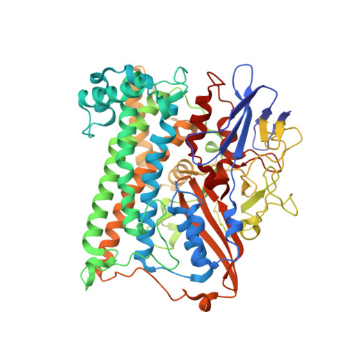
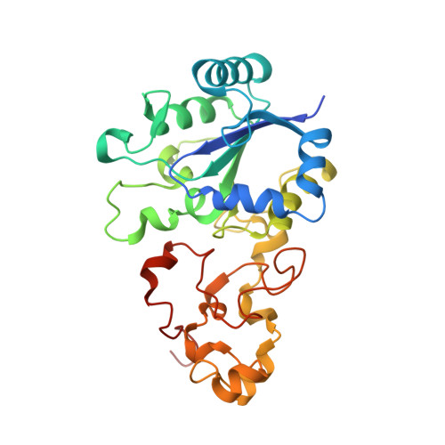
Structural basis for a [4Fe-3S] cluster in the oxygen-tolerant membrane-bound [NiFe]-hydrogenase

Shomura, Y.Yoon, K.S.Nishihara, H.Higuchi, Y.

(2011) Nature 479: 253-256

  • DOI: https://doi.org/10.1038/nature10504
  • Primary Citation of Related Structures:  
    3AYX, 3AYZ, 5Y34

  • PubMed Abstract: 

    Membrane-bound respiratory [NiFe]-hydrogenase (MBH), a H(2)-uptake enzyme found in the periplasmic space of bacteria, catalyses the oxidation of dihydrogen: H(2) → 2H(+) + 2e(-) (ref. 1). In contrast to the well-studied O(2)-sensitive [NiFe]-hydrogenases (referred to as the standard enzymes), MBH has an O(2)-tolerant H(2) oxidation activity; however, the mechanism of O(2) tolerance is unclear. Here we report the crystal structures of Hydrogenovibrio marinus MBH in three different redox conditions at resolutions between 1.18 and 1.32 Å. We find that the proximal iron-sulphur (Fe-S) cluster of MBH has a [4Fe-3S] structure coordinated by six cysteine residues--in contrast to the [4Fe-4S] cubane structure coordinated by four cysteine residues found in the proximal Fe-S cluster of the standard enzymes--and that an amide nitrogen of the polypeptide backbone is deprotonated and additionally coordinates the cluster when chemically oxidized, thus stabilizing the superoxidized state of the cluster. The structure of MBH is very similar to that of the O(2)-sensitive standard enzymes except for the proximal Fe-S cluster. Our results give a reasonable explanation why the O(2) tolerance of MBH is attributable to the unique proximal Fe-S cluster; we propose that the cluster is not only a component of the electron transfer for the catalytic cycle, but that it also donates two electrons and one proton crucial for the appropriate reduction of O(2) in preventing the formation of an unready, inactive state of the enzyme.


  • Organizational Affiliation

    Department of Life Science, Graduate School of Life Science, University of Hyogo, 3-2-1 Koto, Kamigori-cho, Ako-gun, Hyogo 678-1297, Japan.


Macromolecules
Find similar proteins by:  (by identity cutoff)  |  3D Structure
Entity ID: 1
MoleculeChains Sequence LengthOrganismDetailsImage
Hydrogenase
A, C
596Hydrogenovibrio marinusMutation(s): 0 
EC: 1.12.5.1
UniProt
Find proteins for F2Z6J6 (Hydrogenovibrio marinus)
Explore F2Z6J6 
Go to UniProtKB:  F2Z6J6
Entity Groups  
Sequence Clusters30% Identity50% Identity70% Identity90% Identity95% Identity100% Identity
UniProt GroupF2Z6J6
Sequence Annotations
Expand
  • Reference Sequence
Find similar proteins by:  (by identity cutoff)  |  3D Structure
Entity ID: 2
MoleculeChains Sequence LengthOrganismDetailsImage
Membrane-bound hydrogenase small subunit
B, D
283Hydrogenovibrio marinusMutation(s): 0 
UniProt
Find proteins for A0A066ZR71 (Hydrogenovibrio marinus)
Explore A0A066ZR71 
Go to UniProtKB:  A0A066ZR71
Entity Groups  
Sequence Clusters30% Identity50% Identity70% Identity90% Identity95% Identity100% Identity
UniProt GroupA0A066ZR71
Sequence Annotations
Expand
  • Reference Sequence
Small Molecules
Ligands 8 Unique
IDChains Name / Formula / InChI Key2D Diagram3D Interactions
SF4
Query on SF4

Download Ideal Coordinates CCD File 
M [auth B],
V [auth D]
IRON/SULFUR CLUSTER
Fe4 S4
LJBDFODJNLIPKO-UHFFFAOYSA-N
SF3
Query on SF3

Download Ideal Coordinates CCD File 
K [auth B],
T [auth D]
FE4-S3 CLUSTER
Fe4 S3
QQACTBFBZNWJMV-UHFFFAOYSA-N
F3S
Query on F3S

Download Ideal Coordinates CCD File 
L [auth B],
U [auth D]
FE3-S4 CLUSTER
Fe3 S4
FCXHZBQOKRZXKS-UHFFFAOYSA-N
FCO
Query on FCO

Download Ideal Coordinates CCD File 
F [auth A],
P [auth C]
CARBONMONOXIDE-(DICYANO) IRON
C3 Fe N2 O
VBQUCMTXYFMTTE-UHFFFAOYSA-N
GOL
Query on GOL

Download Ideal Coordinates CCD File 
H [auth A],
I [auth A],
R [auth C],
S [auth C]
GLYCEROL
C3 H8 O3
PEDCQBHIVMGVHV-UHFFFAOYSA-N
3NI
Query on 3NI

Download Ideal Coordinates CCD File 
E [auth A],
O [auth C]
NICKEL (III) ION
Ni
JDRCAGKFDGHRNQ-UHFFFAOYSA-N
MG
Query on MG

Download Ideal Coordinates CCD File 
J [auth A],
N [auth C]
MAGNESIUM ION
Mg
JLVVSXFLKOJNIY-UHFFFAOYSA-N
O
Query on O

Download Ideal Coordinates CCD File 
G [auth A],
Q [auth C]
OXYGEN ATOM
O
XLYOFNOQVPJJNP-UHFFFAOYSA-N
Experimental Data & Validation

Experimental Data

  • Method: X-RAY DIFFRACTION
  • Resolution: 1.32 Å
  • R-Value Free: 0.158 
  • R-Value Work: 0.131 
  • R-Value Observed: 0.132 
  • Space Group: P 1 21 1
Unit Cell:
Length ( Å )Angle ( ˚ )
a = 75.628α = 90
b = 116.943β = 91.42
c = 113.337γ = 90
Software Package:
Software NamePurpose
REFMACrefinement
HKL-2000data reduction
HKL-2000data scaling
MOLREPphasing

Structure Validation

View Full Validation Report


Entry History & Funding Information

Deposition Data


Funding OrganizationLocationGrant Number
MEXTJapan20051022
MEXTJapan22770111
MEXTJapan18GS0207
JSPSJapan20580094
JSPSJapan22370061

Revision History  (Full details and data files)

  • Version 1.0: 2017-08-16
    Type: Initial release
  • Version 1.1: 2023-11-22
    Changes: Data collection, Database references, Derived calculations, Refinement description