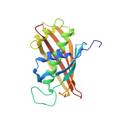
Structural analysis of the dual-function thioesterase SAV606 unravels the mechanism of Michael addition of glycine to an alpha , beta-unsaturated thioester.
Chisuga, T., Miyanaga, A., Kudo, F., Eguchi, T.(2017) J Biol Chem 292: 10926-10937
- PubMed: 28522606
- DOI: https://doi.org/10.1074/jbc.M117.792549
- Primary Citation of Related Structures:
5WSX, 5WSY - PubMed Abstract:
Thioesterases catalyze hydrolysis of acyl thioesters to release carboxylic acid or macrocyclization to produce the corresponding macrocycle in the biosynthesis of fatty acids, polyketides, or nonribosomal peptides. Recently, we reported that the thioesterase CmiS1 from Streptomyces sp. MJ635-86F5 catalyzes the Michael addition of glycine to an α,β-unsaturated fatty acyl thioester followed by thioester hydrolysis in the biosynthesis of the macrolactam antibiotic cremimycin. However, the molecular mechanisms of CmiS1-catalyzed reactions are unclear. Here, we report on the functional and structural characterization of the CmiS1 homolog SAV606 from Streptomyces avermitilis MA-4680. In vitro analysis indicated that SAV606 catalyzes the Michael addition of glycine to crotonic acid thioester and subsequent hydrolysis yielding ( R )- N -carboxymethyl-3-aminobutyric acid. We also determined the crystal structures of SAV606 both in ligand-free form at 2.4 Å resolution and in complex with ( R )- N -carboxymethyl-3-aminobutyric acid at 2.0 Å resolution. We found that SAV606 adopts an α/β hotdog fold and has an active site at the dimeric interface. Examining the complexed structure, we noted that the substrate-binding loop comprising Tyr-53-Asn-61 recognizes the glycine moiety of ( R )- N -carboxymethyl-3-aminobutyric acid. Moreover, we found that SAV606 does not contain an acidic residue at the active site, which is distinct from canonical hotdog thioesterases. Site-directed mutagenesis experiments revealed that His-59 plays a crucial role in both the Michael addition and hydrolysis via a water molecule. These results allow us to propose the reaction mechanism of the SAV606-catalyzed Michael addition and thioester hydrolysis and provide new insight into the multiple functions of a thioesterase family enzyme.
Organizational Affiliation:
From the Department of Chemistry and Materials Science and.