5VCN

THE CRYSTAL STRUCTURE OF DER P 1 ALLERGEN COMPLEXED WITH FAB FRAGMENT OF MAB 5H8


Experimental Data Snapshot

  • Method: X-RAY DIFFRACTION
  • Resolution: 3.00 Å
  • R-Value Free: 0.265 
  • R-Value Work: 0.219 
  • R-Value Observed: 0.221 

wwPDB Validation   3D Report Full Report


Ligand Structure Quality Assessment 


This is version 1.7 of the entry. See complete history


Literature

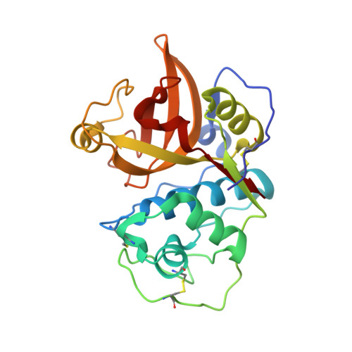
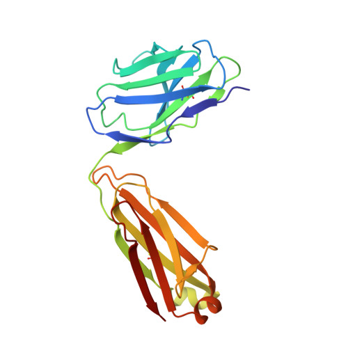
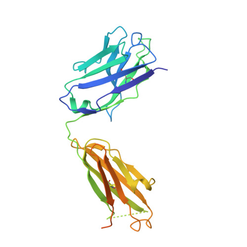
Structural Analysis of Der p 1-Antibody Complexes and Comparison with Complexes of Proteins or Peptides with Monoclonal Antibodies.

Osinski, T.Pomes, A.Majorek, K.A.Glesner, J.Offermann, L.R.Vailes, L.D.Chapman, M.D.Minor, W.Chruszcz, M.

(2015) J Immunol 195: 307-316

  • DOI: https://doi.org/10.4049/jimmunol.1402199
  • Primary Citation of Related Structures:  
    4POZ, 5VCN, 5VCO

  • PubMed Abstract: 

    Der p 1 is a major allergen from the house dust mite, Dermatophagoides pteronyssinus, that belongs to the papain-like cysteine protease family. To investigate the antigenic determinants of Der p 1, we determined two crystal structures of Der p 1 in complex with the Fab fragments of mAbs 5H8 or 10B9. Epitopes for these two Der p 1-specific Abs are located in different, nonoverlapping parts of the Der p 1 molecule. Nevertheless, surface area and identity of the amino acid residues involved in hydrogen bonds between allergen and Ab are similar. The epitope for mAb 10B9 only showed a partial overlap with the previously reported epitope for mAb 4C1, a cross-reactive mAb that binds Der p 1 and its homolog Der f 1 from Dermatophagoides farinae. Upon binding to Der p 1, the Fab fragment of mAb 10B9 was found to form a very rare α helix in its third CDR of the H chain. To provide an overview of the surface properties of the interfaces formed by the complexes of Der p 1-10B9 and Der p 1-5H8, along with the complexes of 4C1 with Der p 1 and Der f 1, a broad analysis of the surfaces and hydrogen bonds of all complexes of Fab-protein or Fab-peptide was performed. This work provides detailed insight into the cross-reactive and specific allergen-Ab interactions in group 1 mite allergens. The surface data of Fab-protein and Fab-peptide interfaces can be used in the design of conformational epitopes with reduced Ab binding for immunotherapy.


  • Organizational Affiliation

    University of Virginia, Charlottesville, VA 22908; Adam Mickiewicz University, 61-712 Poznan, Poland;


Macromolecules
Find similar proteins by:  (by identity cutoff)  |  3D Structure
Entity ID: 1
MoleculeChains Sequence LengthOrganismDetailsImage
PEPTIDASE 1
A, B
222Dermatophagoides pteronyssinusMutation(s): 0 
UniProt
Find proteins for P08176 (Dermatophagoides pteronyssinus)
Explore P08176 
Go to UniProtKB:  P08176
Entity Groups  
Sequence Clusters30% Identity50% Identity70% Identity90% Identity95% Identity100% Identity
UniProt GroupP08176
Sequence Annotations
Expand
  • Reference Sequence
Find similar proteins by:  (by identity cutoff)  |  3D Structure
Entity ID: 2
MoleculeChains Sequence LengthOrganismDetailsImage
LIGHT CHAIN OF FAB FRAGMENT OF MAB 5H8
C, E
210Mus musculusMutation(s): 0 
Entity Groups  
Sequence Clusters30% Identity50% Identity70% Identity90% Identity95% Identity100% Identity
Sequence Annotations
Expand
  • Reference Sequence
Find similar proteins by:  (by identity cutoff)  |  3D Structure
Entity ID: 3
MoleculeChains Sequence LengthOrganismDetailsImage
HEAVY CHAIN OF FAB FRAGMENT OF MAB 5H8
D, F
260Mus musculusMutation(s): 0 
Entity Groups  
Sequence Clusters30% Identity50% Identity70% Identity90% Identity95% Identity100% Identity
Sequence Annotations
Expand
  • Reference Sequence
Small Molecules
Ligands 4 Unique
IDChains Name / Formula / InChI Key2D Diagram3D Interactions
NAG
Query on NAG

Download Ideal Coordinates CCD File 
J [auth A],
N [auth B]
2-acetamido-2-deoxy-beta-D-glucopyranose
C8 H15 N O6
OVRNDRQMDRJTHS-FMDGEEDCSA-N
PO4
Query on PO4

Download Ideal Coordinates CCD File 
H [auth A],
L [auth B]
PHOSPHATE ION
O4 P
NBIIXXVUZAFLBC-UHFFFAOYSA-K
EDO
Query on EDO

Download Ideal Coordinates CCD File 
I [auth A],
M [auth B]
1,2-ETHANEDIOL
C2 H6 O2
LYCAIKOWRPUZTN-UHFFFAOYSA-N
CA
Query on CA

Download Ideal Coordinates CCD File 
G [auth A],
K [auth B]
CALCIUM ION
Ca
BHPQYMZQTOCNFJ-UHFFFAOYSA-N
Experimental Data & Validation

Experimental Data

  • Method: X-RAY DIFFRACTION
  • Resolution: 3.00 Å
  • R-Value Free: 0.265 
  • R-Value Work: 0.219 
  • R-Value Observed: 0.221 
  • Space Group: P 1 21 1
Unit Cell:
Length ( Å )Angle ( ˚ )
a = 47.743α = 90
b = 73.263β = 91.09
c = 200.34γ = 90
Software Package:
Software NamePurpose
HKL-3000data reduction
HKL-3000data scaling
HKL-3000phasing
MOLREPphasing
REFMACrefinement

Structure Validation

View Full Validation Report


Ligand Structure Quality Assessment 


Entry History & Funding Information

Deposition Data


Funding OrganizationLocationGrant Number
National Institutes of Health/National Institute Of Allergy and Infectious Diseases (NIH/NIAID)United StatesR01AI077653
National Institutes of Health/National Institute of General Medical Sciences (NIH/NIGMS)United StatesGM53163

Revision History  (Full details and data files)

  • Version 1.0: 2017-04-26
    Type: Initial release
  • Version 1.1: 2017-05-03
    Changes: Derived calculations
  • Version 1.2: 2017-09-20
    Changes: Author supporting evidence, Refinement description
  • Version 1.3: 2019-12-11
    Changes: Author supporting evidence
  • Version 1.4: 2020-07-29
    Type: Remediation
    Reason: Carbohydrate remediation
    Changes: Data collection, Derived calculations, Structure summary
  • Version 1.5: 2022-04-13
    Changes: Database references, Structure summary
  • Version 1.6: 2022-04-27
    Changes: Data collection
  • Version 1.7: 2023-10-04
    Changes: Data collection, Refinement description