5ULI

Crystal Structure of mouse DXO in complex with (3'-NADP)+ and calcium ion


Experimental Data Snapshot

  • Method: X-RAY DIFFRACTION
  • Resolution: 2.10 Å
  • R-Value Free: 0.208 
  • R-Value Work: 0.161 
  • R-Value Observed: 0.165 

wwPDB Validation   3D Report Full Report


Ligand Structure Quality Assessment 


This is version 1.1 of the entry. See complete history


Literature

5' End Nicotinamide Adenine Dinucleotide Cap in Human Cells Promotes RNA Decay through DXO-Mediated deNADding.

Jiao, X.Doamekpor, S.K.Bird, J.G.Nickels, B.E.Tong, L.Hart, R.P.Kiledjian, M.

(2017) Cell 168: 1015-1027.e10

  • DOI: https://doi.org/10.1016/j.cell.2017.02.019
  • Primary Citation of Related Structures:  
    5ULI, 5ULJ

  • PubMed Abstract: 

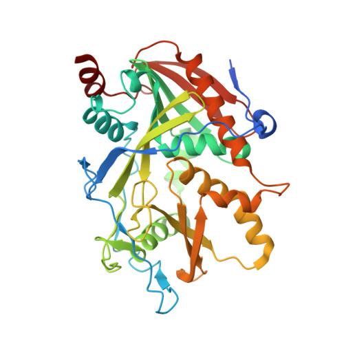
    Eukaryotic mRNAs generally possess a 5' end N7 methyl guanosine (m 7 G) cap that promotes their translation and stability. However, mammalian mRNAs can also carry a 5' end nicotinamide adenine dinucleotide (NAD + ) cap that, in contrast to the m 7 G cap, does not support translation but instead promotes mRNA decay. The mammalian and fungal noncanonical DXO/Rai1 decapping enzymes efficiently remove NAD + caps, and cocrystal structures of DXO/Rai1 with 3'-NADP + illuminate the molecular mechanism for how the "deNADding" reaction produces NAD + and 5' phosphate RNA. Removal of DXO from cells increases NAD + -capped mRNA levels and enables detection of NAD + -capped intronic small nucleolar RNAs (snoRNAs), suggesting NAD + caps can be added to 5'-processed termini. Our findings establish NAD + as an alternative mammalian RNA cap and DXO as a deNADding enzyme modulating cellular levels of NAD + -capped RNAs. Collectively, these data reveal that mammalian RNAs can harbor a 5' end modification distinct from the classical m 7 G cap that promotes rather than inhibits RNA decay.


  • Organizational Affiliation

    Department of Cell Biology and Neuroscience, Rutgers University, Piscataway, NJ 08854, USA.


Macromolecules
Find similar proteins by:  (by identity cutoff)  |  3D Structure
Entity ID: 1
MoleculeChains Sequence LengthOrganismDetailsImage
Decapping and exoribonuclease protein378Mus musculusMutation(s): 0 
Gene Names: DxoDom3zNg6
EC: 3.1.13 (PDB Primary Data), 3.6.1 (PDB Primary Data)
UniProt
Find proteins for O70348 (Mus musculus)
Explore O70348 
Go to UniProtKB:  O70348
Entity Groups  
Sequence Clusters30% Identity50% Identity70% Identity90% Identity95% Identity100% Identity
UniProt GroupO70348
Sequence Annotations
Expand
  • Reference Sequence
Small Molecules
Ligands 2 Unique
IDChains Name / Formula / InChI Key2D Diagram3D Interactions
0WD
Query on 0WD

Download Ideal Coordinates CCD File 
C [auth A][[(2R,3S,4R,5R)-5-(3-aminocarbonyl-4H-pyridin-1-yl)-3,4-bis(oxidanyl)oxolan-2-yl]methoxy-oxidanyl-phosphoryl] [(2R,3S,4R,5R)-5-(6-aminopurin-9-yl)-4-oxidanyl-3-phosphonooxy-oxolan-2-yl]methyl hydrogen phosphate
C21 H30 N7 O17 P3
ONOVTRNLNDQWGA-NNYOXOHSSA-N
CA
Query on CA

Download Ideal Coordinates CCD File 
B [auth A]CALCIUM ION
Ca
BHPQYMZQTOCNFJ-UHFFFAOYSA-N
Experimental Data & Validation

Experimental Data

  • Method: X-RAY DIFFRACTION
  • Resolution: 2.10 Å
  • R-Value Free: 0.208 
  • R-Value Work: 0.161 
  • R-Value Observed: 0.165 
  • Space Group: P 1 21 1
Unit Cell:
Length ( Å )Angle ( ˚ )
a = 49.867α = 90
b = 87.678β = 113.02
c = 53.158γ = 90
Software Package:
Software NamePurpose
PHENIXrefinement
XSCALEdata scaling
PHASERphasing
PDB_EXTRACTdata extraction
XDSdata reduction

Structure Validation

View Full Validation Report


Ligand Structure Quality Assessment 


Entry History 

Deposition Data

Revision History  (Full details and data files)

  • Version 1.0: 2017-05-17
    Type: Initial release
  • Version 1.1: 2024-03-06
    Changes: Data collection, Database references, Derived calculations