5U5I

The dimeric crystal structure of the selenomethionine derivative of HTPA Reductase from Sellaginella moellendorffii


Experimental Data Snapshot

  • Method: X-RAY DIFFRACTION
  • Resolution: 2.20 Å
  • R-Value Free: 0.245 
  • R-Value Work: 0.200 
  • R-Value Observed: 0.203 

wwPDB Validation   3D Report Full Report


Ligand Structure Quality Assessment 


This is version 1.2 of the entry. See complete history


Literature

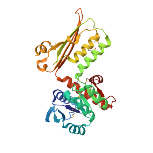
Plant DHDPR forms a dimer with unique secondary structure features that preclude higher-order assembly.

Watkin, S.A.J.Keown, J.R.Richards, E.Goldstone, D.C.Devenish, S.R.A.Grant Pearce, F.

(2018) Biochem J 475: 137-150

  • DOI: https://doi.org/10.1042/BCJ20170709
  • Primary Citation of Related Structures:  
    5U5I, 5U5N, 5UA0, 5UGJ

  • PubMed Abstract: 

    Dihydrodipicolinate reductase (DHDPR) catalyses the second reaction in the diaminopimelate pathway of lysine biosynthesis in bacteria and plants. In contrast with the tetrameric bacterial DHDPR enzymes, we show that DHDPR from Vitis vinifera (grape) and Selaginella moellendorffii are dimeric in solution. In the present study, we have also determined the crystal structures of DHDPR enzymes from the plants Arabidopsis thaliana and S. moellendorffii , which are the first dimeric DHDPR structures. The analysis of these models demonstrates that the dimer forms through the intra-strand interface, and that unique secondary features in the plant enzymes block tetramer assembly. In addition, we have also solved the structure of tetrameric DHDPR from the pathogenic bacteria Neisseria meningitidis Measuring the activity of plant DHDPR enzymes showed that they are much more prone to substrate inhibition than the bacterial enzymes, which appears to be a consequence of increased flexibility of the substrate-binding loop and higher affinity for the nucleotide substrate. This higher propensity to substrate inhibition may have consequences for ongoing efforts to increase lysine biosynthesis in plants.


  • Organizational Affiliation

    Biomolecular Interaction Centre and School of Biological Sciences, University of Canterbury, Christchurch, New Zealand.


Macromolecules
Find similar proteins by:  (by identity cutoff)  |  3D Structure
Entity ID: 1
MoleculeChains Sequence LengthOrganismDetailsImage
HTPA Reductase
A, B
278Selaginella moellendorffiiMutation(s): 0 
Gene Names: SELMODRAFT_168311
UniProt
Find proteins for D8R6G2 (Selaginella moellendorffii)
Explore D8R6G2 
Go to UniProtKB:  D8R6G2
Entity Groups  
Sequence Clusters30% Identity50% Identity70% Identity90% Identity95% Identity100% Identity
UniProt GroupD8R6G2
Sequence Annotations
Expand
  • Reference Sequence
Small Molecules
Modified Residues  1 Unique
IDChains TypeFormula2D DiagramParent
MSE
Query on MSE
A, B
L-PEPTIDE LINKINGC5 H11 N O2 SeMET
Experimental Data & Validation

Experimental Data

  • Method: X-RAY DIFFRACTION
  • Resolution: 2.20 Å
  • R-Value Free: 0.245 
  • R-Value Work: 0.200 
  • R-Value Observed: 0.203 
  • Space Group: P 21 21 21
Unit Cell:
Length ( Å )Angle ( ˚ )
a = 63.922α = 90
b = 64.676β = 90
c = 151.957γ = 90
Software Package:
Software NamePurpose
REFMACrefinement
Aimlessdata scaling
SHELXDEphasing
PDB_EXTRACTdata extraction
XDSdata reduction
SHELXDphasing

Structure Validation

View Full Validation Report


Ligand Structure Quality Assessment 


Entry History 

Deposition Data

Revision History  (Full details and data files)

  • Version 1.0: 2017-12-06
    Type: Initial release
  • Version 1.1: 2017-12-13
    Changes: Database references
  • Version 1.2: 2018-01-17
    Changes: Database references