5TPW

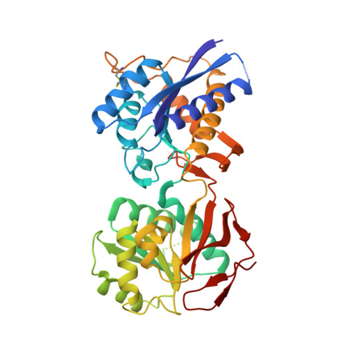
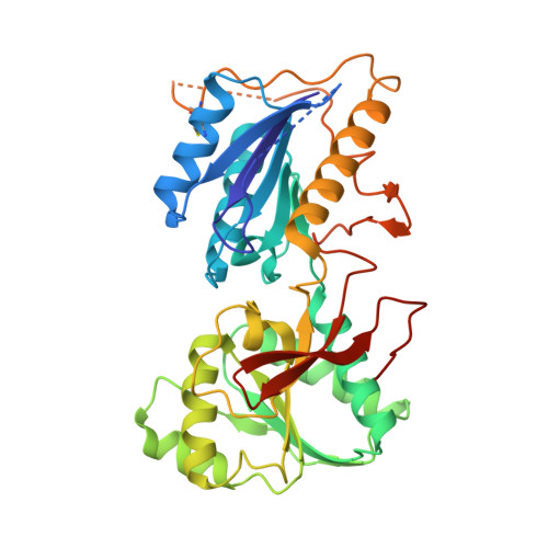
Crystal structure of amino terminal domains of the NMDA receptor subunit GluN1 and GluN2A in complex with zinc at the GluN2A


Experimental Data Snapshot

  • Method: X-RAY DIFFRACTION
  • Resolution: 2.91 Å
  • R-Value Free: 0.245 
  • R-Value Work: 0.200 
  • R-Value Observed: 0.204 

wwPDB Validation   3D Report Full Report


Ligand Structure Quality Assessment 


This is version 1.5 of the entry. See complete history


Literature

Molecular Basis for Subtype Specificity and High-Affinity Zinc Inhibition in the GluN1-GluN2A NMDA Receptor Amino-Terminal Domain.

Romero-Hernandez, A.Simorowski, N.Karakas, E.Furukawa, H.

(2016) Neuron 92: 1324-1336

  • DOI: https://doi.org/10.1016/j.neuron.2016.11.006
  • Primary Citation of Related Structures:  
    5TPW, 5TPZ, 5TQ0, 5TQ2

  • PubMed Abstract: 

    Zinc is vastly present in the mammalian brain and controls functions of various cell surface receptors to regulate neurotransmission. A distinctive characteristic of N-methyl-D-aspartate (NMDA) receptors containing a GluN2A subunit is that their ion channel activity is allosterically inhibited by a nano-molar concentration of zinc that binds to an extracellular domain called an amino-terminal domain (ATD). Despite physiological importance, the molecular mechanism underlying the high-affinity zinc inhibition has been incomplete because of the lack of a GluN2A ATD structure. Here we show the first crystal structures of the heterodimeric GluN1-GluN2A ATD, which provide the complete map of the high-affinity zinc-binding site and reveal distinctive features from the ATD of the GluN1-GluN2B subtype. Perturbation of hydrogen bond networks at the hinge of the GluN2A bi-lobe structure affects both zinc inhibition and open probability, supporting the general model in which the bi-lobe motion in ATD regulates the channel activity in NMDA receptors.


  • Organizational Affiliation

    WM Keck Structural Biology Laboratory, Cold Spring Harbor Laboratory, Cold Spring Harbor, NY 11724, USA; Watson School of Biological Sciences, Cold Spring Harbor, NY 11724, USA.


Macromolecules
Find similar proteins by:  (by identity cutoff)  |  3D Structure
Entity ID: 1
MoleculeChains Sequence LengthOrganismDetailsImage
NMDA glutamate receptor subunit389Xenopus laevisMutation(s): 2 
Gene Names: grin1NR1
UniProt
Find proteins for A0A1L8F5J9 (Xenopus laevis)
Explore A0A1L8F5J9 
Go to UniProtKB:  A0A1L8F5J9
Entity Groups  
Sequence Clusters30% Identity50% Identity70% Identity90% Identity95% Identity100% Identity
UniProt GroupA0A1L8F5J9
Sequence Annotations
Expand
  • Reference Sequence
Find similar proteins by:  (by identity cutoff)  |  3D Structure
Entity ID: 2
MoleculeChains Sequence LengthOrganismDetailsImage
Glutamate receptor ionotropic, NMDA 2A360Rattus norvegicusMutation(s): 0 
Gene Names: Grin2a
UniProt
Find proteins for Q00959 (Rattus norvegicus)
Explore Q00959 
Go to UniProtKB:  Q00959
Entity Groups  
Sequence Clusters30% Identity50% Identity70% Identity90% Identity95% Identity100% Identity
UniProt GroupQ00959
Sequence Annotations
Expand
  • Reference Sequence
Find similar proteins by:  (by identity cutoff)  |  3D Structure
Entity ID: 3
MoleculeChains Sequence LengthOrganismDetailsImage
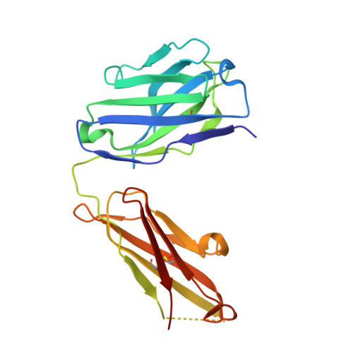
FAB, HEAVY CHAINC [auth H]221Mus musculusMutation(s): 0 
Entity Groups  
Sequence Clusters30% Identity50% Identity70% Identity90% Identity95% Identity100% Identity
Sequence Annotations
Expand
  • Reference Sequence
Find similar proteins by:  (by identity cutoff)  |  3D Structure
Entity ID: 4
MoleculeChains Sequence LengthOrganismDetailsImage
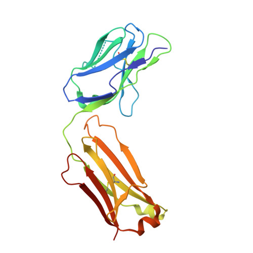
FAB, LIGHT CHAIND [auth L]214Mus musculusMutation(s): 0 
Entity Groups  
Sequence Clusters30% Identity50% Identity70% Identity90% Identity95% Identity100% Identity
Sequence Annotations
Expand
  • Reference Sequence
Small Molecules
Ligands 4 Unique
IDChains Name / Formula / InChI Key2D Diagram3D Interactions
NAG
Query on NAG

Download Ideal Coordinates CCD File 
E [auth A],
F [auth A]
2-acetamido-2-deoxy-beta-D-glucopyranose
C8 H15 N O6
OVRNDRQMDRJTHS-FMDGEEDCSA-N
SO4
Query on SO4

Download Ideal Coordinates CCD File 
G [auth A]
H [auth A]
I [auth A]
M [auth B]
N [auth B]
G [auth A],
H [auth A],
I [auth A],
M [auth B],
N [auth B],
O [auth B]
SULFATE ION
O4 S
QAOWNCQODCNURD-UHFFFAOYSA-L
GOL
Query on GOL

Download Ideal Coordinates CCD File 
J [auth A],
K [auth A]
GLYCEROL
C3 H8 O3
PEDCQBHIVMGVHV-UHFFFAOYSA-N
ZN
Query on ZN

Download Ideal Coordinates CCD File 
L [auth B]ZINC ION
Zn
PTFCDOFLOPIGGS-UHFFFAOYSA-N
Experimental Data & Validation

Experimental Data

  • Method: X-RAY DIFFRACTION
  • Resolution: 2.91 Å
  • R-Value Free: 0.245 
  • R-Value Work: 0.200 
  • R-Value Observed: 0.204 
  • Space Group: P 21 21 21
Unit Cell:
Length ( Å )Angle ( ˚ )
a = 79.56α = 90
b = 118.401β = 90
c = 155.812γ = 90
Software Package:
Software NamePurpose
PHENIXrefinement
HKL-2000data collection
HKL-2000data reduction
PHENIXmodel building
PDB_EXTRACTdata extraction
PHASERphasing
DENZOdata reduction
SCALEPACKdata scaling

Structure Validation

View Full Validation Report


Ligand Structure Quality Assessment 


Entry History & Funding Information

Deposition Data


Funding OrganizationLocationGrant Number
National Institutes of Health/National Institute of Mental Health (NIH/NIMH)United StatesMH085926
National Institutes of Health/National Institute of General Medical Sciences (NIH/NIGMS)United StatesGM105730

Revision History  (Full details and data files)

  • Version 1.0: 2016-12-14
    Type: Initial release
  • Version 1.1: 2017-09-20
    Changes: Author supporting evidence, Refinement description
  • Version 1.2: 2018-05-16
    Changes: Data collection, Database references
  • Version 1.3: 2019-11-27
    Changes: Author supporting evidence
  • Version 1.4: 2020-07-29
    Type: Remediation
    Reason: Carbohydrate remediation
    Changes: Data collection, Derived calculations, Structure summary
  • Version 1.5: 2023-10-04
    Changes: Data collection, Database references, Refinement description, Structure summary