5T9T

Protocadherin Gamma B2 extracellular cadherin domains 1-5


Experimental Data Snapshot

  • Method: X-RAY DIFFRACTION
  • Resolution: 3.50 Å
  • R-Value Free: 0.257 
  • R-Value Work: 0.213 
  • R-Value Observed: 0.215 

wwPDB Validation   3D Report Full Report


Ligand Structure Quality Assessment 


This is version 2.2 of the entry. See complete history


Literature

gamma-Protocadherin structural diversity and functional implications.

Goodman, K.M.Rubinstein, R.Thu, C.A.Mannepalli, S.Bahna, F.Ahlsen, G.Rittenhouse, C.Maniatis, T.Honig, B.Shapiro, L.

(2016) Elife 5

  • DOI: https://doi.org/10.7554/eLife.20930
  • Primary Citation of Related Structures:  
    5SZL, 5SZM, 5SZN, 5SZO, 5SZP, 5SZQ, 5SZR, 5T9T

  • PubMed Abstract: 

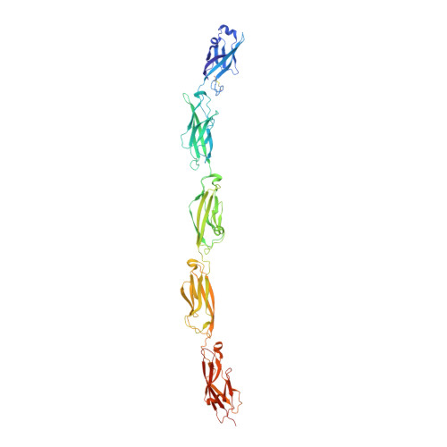
    Stochastic cell-surface expression of α-, β-, and γ-clustered protocadherins (Pcdhs) provides vertebrate neurons with single-cell identities that underlie neuronal self-recognition. Here we report crystal structures of ectodomain fragments comprising cell-cell recognition regions of mouse γ-Pcdhs γA1, γA8, γB2, and γB7 revealing trans -homodimers, and of C-terminal ectodomain fragments from γ-Pcdhs γA4 and γB2, which depict cis -interacting regions in monomeric form. Together these structures span the entire γ-Pcdh ectodomain. The trans -dimer structures reveal determinants of γ-Pcdh isoform-specific homophilic recognition. We identified and structurally mapped cis -dimerization mutations to the C-terminal ectodomain structures. Biophysical studies showed that Pcdh ectodomains from γB-subfamily isoforms formed cis dimers, whereas γA isoforms did not, but both γA and γB isoforms could interact in cis with α-Pcdhs. Together, these data show how interaction specificity is distributed over all domains of the γ-Pcdh trans interface, and suggest that subfamily- or isoform-specific cis -interactions may play a role in the Pcdh-mediated neuronal self-recognition code.


  • Organizational Affiliation

    Department of Biochemistry and Molecular Biophysics, Columbia University, New York, United States.


Macromolecules
Find similar proteins by:  (by identity cutoff)  |  3D Structure
Entity ID: 1
MoleculeChains Sequence LengthOrganismDetailsImage
Protocadherin gamma B2-alpha C
A, B
535Mus musculusMutation(s): 0 
UniProt
Find proteins for Q8K486 (Mus musculus)
Explore Q8K486 
Go to UniProtKB:  Q8K486
Entity Groups  
Sequence Clusters30% Identity50% Identity70% Identity90% Identity95% Identity100% Identity
UniProt GroupQ8K486
Sequence Annotations
Expand
  • Reference Sequence
Oligosaccharides

Help

Entity ID: 2
MoleculeChains Length2D Diagram Glycosylation3D Interactions
beta-D-mannopyranose-(1-4)-2-acetamido-2-deoxy-beta-D-glucopyranose-(1-4)-[alpha-L-fucopyranose-(1-6)]2-acetamido-2-deoxy-beta-D-glucopyranose
C
4N-Glycosylation
Glycosylation Resources
GlyTouCan:  G32152BH
GlyCosmos:  G32152BH
GlyGen:  G32152BH
Small Molecules
Ligands 3 Unique
IDChains Name / Formula / InChI Key2D Diagram3D Interactions
NAG
Query on NAG

Download Ideal Coordinates CCD File 
QA [auth B],
R [auth A],
T [auth A]
2-acetamido-2-deoxy-beta-D-glucopyranose
C8 H15 N O6
OVRNDRQMDRJTHS-FMDGEEDCSA-N
MAN
Query on MAN

Download Ideal Coordinates CCD File 
KA [auth B]
LA [auth B]
MA [auth B]
NA [auth B]
OA [auth B]
KA [auth B],
LA [auth B],
MA [auth B],
NA [auth B],
OA [auth B],
P [auth A],
PA [auth B],
Q [auth A],
S [auth A],
U [auth A],
V [auth A]
alpha-D-mannopyranose
C6 H12 O6
WQZGKKKJIJFFOK-PQMKYFCFSA-N
CA
Query on CA

Download Ideal Coordinates CCD File 
AA [auth B]
BA [auth B]
CA [auth B]
D [auth A]
DA [auth B]
AA [auth B],
BA [auth B],
CA [auth B],
D [auth A],
DA [auth B],
E [auth A],
EA [auth B],
F [auth A],
FA [auth B],
G [auth A],
GA [auth B],
H [auth A],
HA [auth B],
I [auth A],
IA [auth B],
J [auth A],
JA [auth B],
K [auth A],
L [auth A],
M [auth A],
N [auth A],
O [auth A],
W [auth B],
X [auth B],
Y [auth B],
Z [auth B]
CALCIUM ION
Ca
BHPQYMZQTOCNFJ-UHFFFAOYSA-N
Experimental Data & Validation

Experimental Data

Unit Cell:
Length ( Å )Angle ( ˚ )
a = 80.722α = 90
b = 118.067β = 103.91
c = 98.19γ = 90
Software Package:
Software NamePurpose
PHENIXrefinement
XDSdata reduction
Aimlessdata scaling
PHASERphasing

Structure Validation

View Full Validation Report


Ligand Structure Quality Assessment 


Entry History & Funding Information

Deposition Data


Funding OrganizationLocationGrant Number
National Institutes of Health/National Institute of General Medical Sciences (NIH/NIGMS)United StatesGM103403
National Institutes of Health/Office of the DirectorUnited StatesOD012351
National Institutes of Health/Office of the DirectorUnited StatesOD021764

Revision History  (Full details and data files)

  • Version 1.0: 2016-10-19
    Type: Initial release
  • Version 1.1: 2016-11-09
    Changes: Database references
  • Version 1.2: 2017-11-01
    Changes: Author supporting evidence, Derived calculations
  • Version 2.0: 2020-07-29
    Type: Remediation
    Reason: Carbohydrate remediation
    Changes: Atomic model, Data collection, Derived calculations, Structure summary
  • Version 2.1: 2022-03-23
    Changes: Author supporting evidence, Database references, Derived calculations, Structure summary
  • Version 2.2: 2023-10-04
    Changes: Data collection, Refinement description