5NO7

Crystal Structure of a Xylan-active Lytic Polysaccharide Monooxygenase from Pycnoporus coccineus.


Experimental Data Snapshot

  • Method: X-RAY DIFFRACTION
  • Resolution: 3.01 Å
  • R-Value Free: 0.240 
  • R-Value Work: 0.230 
  • R-Value Observed: 0.231 

wwPDB Validation   3D Report Full Report


This is version 2.0 of the entry. See complete history


Literature

Lytic xylan oxidases from wood-decay fungi unlock biomass degradation.

Couturier, M.Ladeveze, S.Sulzenbacher, G.Ciano, L.Fanuel, M.Moreau, C.Villares, A.Cathala, B.Chaspoul, F.Frandsen, K.E.Labourel, A.Herpoel-Gimbert, I.Grisel, S.Haon, M.Lenfant, N.Rogniaux, H.Ropartz, D.Davies, G.J.Rosso, M.N.Walton, P.H.Henrissat, B.Berrin, J.G.

(2018) Nat Chem Biol 14: 306-310

  • DOI: https://doi.org/10.1038/nchembio.2558
  • Primary Citation of Related Structures:  
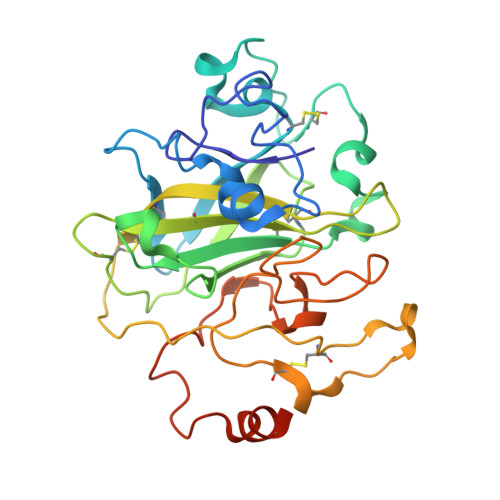
    5NO7

  • PubMed Abstract: 

    Wood biomass is the most abundant feedstock envisioned for the development of modern biorefineries. However, the cost-effective conversion of this form of biomass into commodity products is limited by its resistance to enzymatic degradation. Here we describe a new family of fungal lytic polysaccharide monooxygenases (LPMOs) prevalent among white-rot and brown-rot basidiomycetes that is active on xylans-a recalcitrant polysaccharide abundant in wood biomass. Two AA14 LPMO members from the white-rot fungus Pycnoporus coccineus substantially increase the efficiency of wood saccharification through oxidative cleavage of highly refractory xylan-coated cellulose fibers. The discovery of this unique enzyme activity advances our knowledge on the degradation of woody biomass in nature and offers an innovative solution for improving enzyme cocktails for biorefinery applications.


  • Organizational Affiliation

    INRA, Aix Marseille University, Biodiversité et Biotechnologie Fongiques (BBF), Marseille, France.


Macromolecules
Find similar proteins by:  (by identity cutoff)  |  3D Structure
Entity ID: 1
MoleculeChains Sequence LengthOrganismDetailsImage
Lytic polysaccharide monooxygenase
A, B
292Trametes cinnabarinaMutation(s): 0 
Gene Names: BN946_scf184298.g17
UniProt
Find proteins for A0A060SRI5 (Pycnoporus cinnabarinus)
Explore A0A060SRI5 
Go to UniProtKB:  A0A060SRI5
Entity Groups  
Sequence Clusters30% Identity50% Identity70% Identity90% Identity95% Identity100% Identity
UniProt GroupA0A060SRI5
Sequence Annotations
Expand
  • Reference Sequence
Oligosaccharides

Help

Entity ID: 2
MoleculeChains Length2D Diagram Glycosylation3D Interactions
beta-D-mannopyranose-(1-4)-2-acetamido-2-deoxy-beta-D-glucopyranose-(1-4)-2-acetamido-2-deoxy-beta-D-glucopyranose
C, H
3N-Glycosylation
Glycosylation Resources
GlyTouCan:  G15407YE
GlyCosmos:  G15407YE
GlyGen:  G15407YE
Entity ID: 3
MoleculeChains Length2D Diagram Glycosylation3D Interactions
alpha-D-mannopyranose-(1-3)-alpha-D-mannopyranose-(1-6)-[alpha-D-mannopyranose-(1-3)]beta-D-mannopyranose-(1-4)-2-acetamido-2-deoxy-beta-D-glucopyranose-(1-4)-2-acetamido-2-deoxy-beta-D-glucopyranose
D
6N-Glycosylation
Glycosylation Resources
GlyTouCan:  G09724ZC
GlyCosmos:  G09724ZC
GlyGen:  G09724ZC
Entity ID: 4
MoleculeChains Length2D Diagram Glycosylation3D Interactions
alpha-D-mannopyranose-(1-3)-[alpha-D-mannopyranose-(1-6)]beta-D-mannopyranose-(1-4)-2-acetamido-2-deoxy-beta-D-glucopyranose-(1-4)-2-acetamido-2-deoxy-beta-D-glucopyranose
E
5N-Glycosylation
Glycosylation Resources
GlyTouCan:  G22768VO
GlyCosmos:  G22768VO
GlyGen:  G22768VO
Entity ID: 5
MoleculeChains Length2D Diagram Glycosylation3D Interactions
alpha-D-mannopyranose-(1-3)-beta-D-mannopyranose-(1-4)-2-acetamido-2-deoxy-beta-D-glucopyranose-(1-4)-2-acetamido-2-deoxy-beta-D-glucopyranose
F
4N-Glycosylation
Glycosylation Resources
GlyTouCan:  G81315DD
GlyCosmos:  G81315DD
GlyGen:  G81315DD
Entity ID: 6
MoleculeChains Length2D Diagram Glycosylation3D Interactions
alpha-D-mannopyranose-(1-2)-[alpha-D-mannopyranose-(1-6)]alpha-D-mannopyranose-(1-3)-[alpha-D-mannopyranose-(1-6)]beta-D-mannopyranose-(1-4)-2-acetamido-2-deoxy-beta-D-glucopyranose-(1-4)-2-acetamido-2-deoxy-beta-D-glucopyranose
G, L
7N-Glycosylation
Glycosylation Resources
GlyTouCan:  G97616MU
GlyCosmos:  G97616MU
GlyGen:  G97616MU
Entity ID: 7
MoleculeChains Length2D Diagram Glycosylation3D Interactions
2-acetamido-2-deoxy-beta-D-glucopyranose-(1-4)-2-acetamido-2-deoxy-beta-D-glucopyranose
I, J, K
2N-Glycosylation
Glycosylation Resources
GlyTouCan:  G42666HT
GlyCosmos:  G42666HT
GlyGen:  G42666HT
Small Molecules
Ligands 1 Unique
IDChains Name / Formula / InChI Key2D Diagram3D Interactions
SO4
Query on SO4

Download Ideal Coordinates CCD File 
M [auth A]
N [auth A]
O [auth A]
P [auth A]
Q [auth A]
M [auth A],
N [auth A],
O [auth A],
P [auth A],
Q [auth A],
R [auth A],
S [auth A],
T [auth B],
U [auth B],
V [auth B],
W [auth B]
SULFATE ION
O4 S
QAOWNCQODCNURD-UHFFFAOYSA-L
Experimental Data & Validation

Experimental Data

  • Method: X-RAY DIFFRACTION
  • Resolution: 3.01 Å
  • R-Value Free: 0.240 
  • R-Value Work: 0.230 
  • R-Value Observed: 0.231 
  • Space Group: P 41 21 2
Unit Cell:
Length ( Å )Angle ( ˚ )
a = 203.754α = 90
b = 203.754β = 90
c = 110.586γ = 90
Software Package:
Software NamePurpose
REFMACrefinement
XDSdata reduction
XDSdata scaling
SHELXCDphasing

Structure Validation

View Full Validation Report


Entry History & Funding Information

Deposition Data


Funding OrganizationLocationGrant Number
French National Research AgencyFranceANR-11-IDEX-0001-02
French National Research AgencyFranceANR-10-INSB-05-01

Revision History  (Full details and data files)

  • Version 1.0: 2018-01-17
    Type: Initial release
  • Version 1.1: 2018-02-07
    Changes: Database references, Structure summary
  • Version 1.2: 2018-02-21
    Changes: Database references
  • Version 2.0: 2020-07-29
    Type: Remediation
    Reason: Carbohydrate remediation
    Changes: Atomic model, Data collection, Derived calculations, Structure summary