5MW1

cryoEM structure of crenactin double helical filament at 3.8A resolution


Experimental Data Snapshot

  • Method: ELECTRON MICROSCOPY
  • Resolution: 3.80 Å
  • Aggregation State: HELICAL ARRAY 
  • Reconstruction Method: HELICAL 

wwPDB Validation   3D Report Full Report


This is version 1.2 of the entry. See complete history


Literature

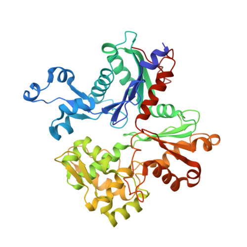
Crenactin forms actin-like double helical filaments regulated by arcadin-2.

Izore, T.Kureisaite-Ciziene, D.McLaughlin, S.H.Lowe, J.

(2016) Elife 5

  • DOI: https://doi.org/10.7554/eLife.21600
  • Primary Citation of Related Structures:  
    5LY3, 5LY5, 5MW1

  • PubMed Abstract: 

    The similarity of eukaryotic actin to crenactin, a filament-forming protein from the crenarchaeon Pyrobaculum calidifontis supports the theory of a common origin of Crenarchaea and Eukaryotes. Monomeric structures of crenactin and actin are similar, although their filament architectures were suggested to be different. Here we report that crenactin forms bona fide double helical filaments that show exceptional similarity to eukaryotic F-actin. With cryo-electron microscopy and helical reconstruction we solved the structure of the crenactin filament to 3.8 Å resolution. When forming double filaments, the 'hydrophobic plug' loop in crenactin rearranges. Arcadin-2, also encoded by the arcade gene cluster, binds tightly with its C-terminus to the hydrophobic groove of crenactin. Binding is reminiscent of eukaryotic actin modulators such as cofilin and thymosin β4 and arcadin-2 is a depolymeriser of crenactin filaments. Our work further supports the theory of shared ancestry of Eukaryotes and Crenarchaea.


  • Organizational Affiliation

    MRC Laboratory of Molecular Biology, Francis Crick Avenue, Cambridge, United Kingdom.


Macromolecules
Find similar proteins by:  (by identity cutoff)  |  3D Structure
Entity ID: 1
MoleculeChains Sequence LengthOrganismDetailsImage
Actin/actin family protein432Pyrobaculum calidifontisMutation(s): 0 
Gene Names: Pcal_1635
UniProt
Find proteins for A3MWN5 (Pyrobaculum calidifontis (strain DSM 21063 / JCM 11548 / VA1))
Explore A3MWN5 
Go to UniProtKB:  A3MWN5
Entity Groups  
Sequence Clusters30% Identity50% Identity70% Identity90% Identity95% Identity100% Identity
UniProt GroupA3MWN5
Sequence Annotations
Expand
  • Reference Sequence
Experimental Data & Validation

Experimental Data

  • Method: ELECTRON MICROSCOPY
  • Resolution: 3.80 Å
  • Aggregation State: HELICAL ARRAY 
  • Reconstruction Method: HELICAL 

Structure Validation

View Full Validation Report


Entry History & Funding Information

Deposition Data


Funding OrganizationLocationGrant Number
Medical Research Council (United Kingdom)United KingdomU105184326
Wellcome TrustUnited Kingdom095514/Z/11/Z

Revision History  (Full details and data files)

  • Version 1.0: 2017-02-08
    Type: Initial release
  • Version 1.1: 2017-09-13
    Changes: Author supporting evidence, Data collection
  • Version 1.2: 2019-10-23
    Changes: Data collection, Other