5LI3

Crystal structure of HDAC-like protein from P. aeruginosa in complex with a photo-switchable inhibitor.


Experimental Data Snapshot

  • Method: X-RAY DIFFRACTION
  • Resolution: 2.40 Å
  • R-Value Free: 0.270 
  • R-Value Work: 0.225 
  • R-Value Observed: 0.227 

wwPDB Validation   3D Report Full Report


Ligand Structure Quality Assessment 


This is version 1.3 of the entry. See complete history


Literature

Toward Photopharmacological Antimicrobial Chemotherapy Using Photoswitchable Amidohydrolase Inhibitors.

Weston, C.E.Kramer, A.Colin, F.Yildiz, O.Baud, M.G.Meyer-Almes, F.J.Fuchter, M.J.

(2017) ACS Infect Dis 3: 152-161

  • DOI: https://doi.org/10.1021/acsinfecdis.6b00148
  • Primary Citation of Related Structures:  
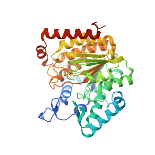
    5G1C, 5G3W, 5LI3

  • PubMed Abstract: 

    Photopharmacological agents exhibit light-dependent biological activity and may have potential in the development of new antimicrobial agents/modalities. Amidohydrolase enzymes homologous to the well-known human histone deacetylases (HDACs) are present in bacteria, including resistant organisms responsible for a significant number of hospital-acquired infections and deaths. We report photopharmacological inhibitors of these enzymes, using two classes of photoswitches embedded in the inhibitor pharmacophore: azobenzenes and arylazopyrazoles. Although both classes of inhibitor show excellent inhibitory activity (nM IC 50 values) of the target enzymes and promising differential activity of the switchable E- and Z-isomeric forms, the arylazopyrazoles exhibit better intrinsic photoswitch performance (more complete switching, longer thermal lifetime of the Z-isomer). We also report protein-ligand crystal structures of the E-isomers of both an azobenzene and an arylazopyrazole inhibitor, bound to bacterial histone deacetylase-like amidohydrolases (HDAHs). These structures not only uncover interactions important for inhibitor binding but also reveal conformational differences between the two photoswitch inhibitor classes. As such, our data may pave the way for the design of improved photopharmacological agents targeting the HDAC superfamily.


  • Organizational Affiliation

    Department of Chemistry, Imperial College London , London SW7 2AZ, United Kingdom.


Macromolecules
Find similar proteins by:  (by identity cutoff)  |  3D Structure
Entity ID: 1
MoleculeChains Sequence LengthOrganismDetailsImage
Acetoin utilization protein
A, B
380Pseudomonas aeruginosaMutation(s): 0 
Gene Names: AO946_14160
UniProt
Find proteins for Q9HXM1 (Pseudomonas aeruginosa (strain ATCC 15692 / DSM 22644 / CIP 104116 / JCM 14847 / LMG 12228 / 1C / PRS 101 / PAO1))
Explore Q9HXM1 
Go to UniProtKB:  Q9HXM1
Entity Groups  
Sequence Clusters30% Identity50% Identity70% Identity90% Identity95% Identity100% Identity
UniProt GroupQ9HXM1
Sequence Annotations
Expand
  • Reference Sequence
Small Molecules
Ligands 3 Unique
IDChains Name / Formula / InChI Key2D Diagram3D Interactions
9RB
Query on 9RB

Download Ideal Coordinates CCD File 
F [auth A],
J [auth B]
(2E)-N-hydroxy-3-{4-[(E)-(1,3,5-trimethyl-1H-pyrazol-4-yl)diazenyl]phenyl}prop-2-enamide
C15 H17 N5 O2
GGZOJTXQALEQJU-OUWLFTCQSA-N
ZN
Query on ZN

Download Ideal Coordinates CCD File 
C [auth A],
G [auth B]
ZINC ION
Zn
PTFCDOFLOPIGGS-UHFFFAOYSA-N
K
Query on K

Download Ideal Coordinates CCD File 
D [auth A],
E [auth A],
H [auth B],
I [auth B]
POTASSIUM ION
K
NPYPAHLBTDXSSS-UHFFFAOYSA-N
Binding Affinity Annotations 
IDSourceBinding Affinity
9RB Binding MOAD:  5LI3 IC50: 53 (nM) from 1 assay(s)
Experimental Data & Validation

Experimental Data

  • Method: X-RAY DIFFRACTION
  • Resolution: 2.40 Å
  • R-Value Free: 0.270 
  • R-Value Work: 0.225 
  • R-Value Observed: 0.227 
  • Space Group: P 41 21 2
Unit Cell:
Length ( Å )Angle ( ˚ )
a = 82.19α = 90
b = 82.19β = 90
c = 217.03γ = 90
Software Package:
Software NamePurpose
REFMACrefinement
MOSFLMdata reduction
Aimlessdata scaling
MOLREPphasing

Structure Validation

View Full Validation Report


Ligand Structure Quality Assessment 


Entry History & Funding Information

Deposition Data


Funding OrganizationLocationGrant Number
German Research FoundationGermanyME 3122/2-1

Revision History  (Full details and data files)

  • Version 1.0: 2016-11-23
    Type: Initial release
  • Version 1.1: 2017-02-22
    Changes: Database references
  • Version 1.2: 2017-09-06
    Changes: Author supporting evidence
  • Version 1.3: 2024-01-10
    Changes: Data collection, Database references, Derived calculations, Refinement description