5L6I

Uba1 in complex with Ub-MLN4924 covalent adduct


Experimental Data Snapshot

  • Method: X-RAY DIFFRACTION
  • Resolution: 2.76 Å
  • R-Value Free: 0.220 
  • R-Value Work: 0.174 
  • R-Value Observed: 0.176 

wwPDB Validation   3D Report Full Report


Ligand Structure Quality Assessment 


This is version 1.4 of the entry. See complete history


Literature

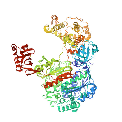
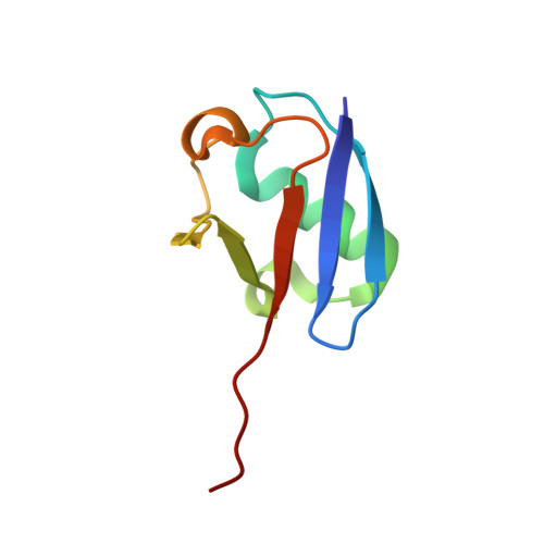
Dissecting the Specificity of Adenosyl Sulfamate Inhibitors Targeting the Ubiquitin-Activating Enzyme.

Misra, M.Kuhn, M.Lobel, M.An, H.Statsyuk, A.V.Sotriffer, C.Schindelin, H.

(2017) Structure 25: 1120-1129.e3

  • DOI: https://doi.org/10.1016/j.str.2017.05.001
  • Primary Citation of Related Structures:  
    5L6H, 5L6I, 5L6J

  • PubMed Abstract: 

    Targeting the activating enzymes (E1) of ubiquitin (Ub) and ubiquitin-like modifiers (Ubls) has emerged as a promising anti-cancer strategy, possibly overcoming the ineffectiveness of proteasome inhibitors against solid tumors. Here, we report crystal structures of the yeast ubiquitin E1 (Uba1) with three adenosyl sulfamate inhibitors exhibiting different E1 specificities, which are all covalently linked to ubiquitin. The structures illustrate how the chemically diverse inhibitors are accommodated within the adenylation active site. When compared with the previously reported structures of various E1 enzymes, our structures provide the basis of the preferences of these inhibitors for different Ub/Ubl-activating enzymes. In vitro inhibition assays and molecular dynamics simulations validated the specificities of the inhibitors as deduced from the structures. Taken together, the structures establish a framework for the development of additional compounds targeting E1 enzymes, which will display higher potency and selectivity.


  • Organizational Affiliation

    Institute of Structural Biology, Rudolf Virchow Center for Experimental Biomedicine, University of Würzburg, Josef-Schneider-Straße 2, 97080 Würzburg, Germany; Institute of Pharmacy and Food Chemistry, Department of Chemistry and Pharmacy, University of Würzburg, Am Hubland, 97074 Würzburg, Germany.


Macromolecules
Find similar proteins by:  (by identity cutoff)  |  3D Structure
Entity ID: 1
MoleculeChains Sequence LengthOrganismDetailsImage
Ubiquitin-activating enzyme E1 1
A, C
1,024Saccharomyces cerevisiae S288CMutation(s): 0 
Gene Names: UBA1YKL210W
EC: 6.2.1.45
UniProt
Find proteins for P22515 (Saccharomyces cerevisiae (strain ATCC 204508 / S288c))
Explore P22515 
Go to UniProtKB:  P22515
Entity Groups  
Sequence Clusters30% Identity50% Identity70% Identity90% Identity95% Identity100% Identity
UniProt GroupP22515
Sequence Annotations
Expand
  • Reference Sequence
Find similar proteins by:  (by identity cutoff)  |  3D Structure
Entity ID: 2
MoleculeChains Sequence LengthOrganismDetailsImage
Ubiquitin-40S ribosomal protein S31
B, D, E
76Saccharomyces cerevisiaeMutation(s): 0 
Gene Names: RPS31RPS37UBI3YLR167WL9470.14
UniProt
Find proteins for P05759 (Saccharomyces cerevisiae (strain ATCC 204508 / S288c))
Explore P05759 
Go to UniProtKB:  P05759
Entity Groups  
Sequence Clusters30% Identity50% Identity70% Identity90% Identity95% Identity100% Identity
UniProt GroupP05759
Sequence Annotations
Expand
  • Reference Sequence
Small Molecules
Ligands 4 Unique
IDChains Name / Formula / InChI Key2D Diagram3D Interactions
B39
Query on B39

Download Ideal Coordinates CCD File 
OA [auth D],
W [auth B]
[(1S,2S,4R)-4-{4-[(1S)-2,3-dihydro-1H-inden-1-ylamino]-7H-pyrrolo[2,3-d]pyrimidin-7-yl}-2-hydroxycyclopentyl]methyl sulfamate
C21 H25 N5 O4 S
MPUQHZXIXSTTDU-QXGSTGNESA-N
SO4
Query on SO4

Download Ideal Coordinates CCD File 
F [auth A],
Y [auth C]
SULFATE ION
O4 S
QAOWNCQODCNURD-UHFFFAOYSA-L
GOL
Query on GOL

Download Ideal Coordinates CCD File 
HA [auth C]
IA [auth C]
JA [auth C]
KA [auth C]
LA [auth C]
HA [auth C],
IA [auth C],
JA [auth C],
KA [auth C],
LA [auth C],
MA [auth C],
N [auth A],
NA [auth C],
O [auth A],
P [auth A],
Q [auth A],
R [auth A],
S [auth A],
T [auth A],
U [auth A],
V [auth A]
GLYCEROL
C3 H8 O3
PEDCQBHIVMGVHV-UHFFFAOYSA-N
CL
Query on CL

Download Ideal Coordinates CCD File 
AA [auth C]
BA [auth C]
CA [auth C]
DA [auth C]
EA [auth C]
AA [auth C],
BA [auth C],
CA [auth C],
DA [auth C],
EA [auth C],
FA [auth C],
G [auth A],
GA [auth C],
H [auth A],
I [auth A],
J [auth A],
K [auth A],
L [auth A],
M [auth A],
X [auth B],
Z [auth C]
CHLORIDE ION
Cl
VEXZGXHMUGYJMC-UHFFFAOYSA-M
Modified Residues  1 Unique
IDChains TypeFormula2D DiagramParent
CSO
Query on CSO
A, C
L-PEPTIDE LINKINGC3 H7 N O3 SCYS
Experimental Data & Validation

Experimental Data

  • Method: X-RAY DIFFRACTION
  • Resolution: 2.76 Å
  • R-Value Free: 0.220 
  • R-Value Work: 0.174 
  • R-Value Observed: 0.176 
  • Space Group: P 2 21 21
Unit Cell:
Length ( Å )Angle ( ˚ )
a = 72.5α = 90
b = 191.89β = 90
c = 230.197γ = 90
Software Package:
Software NamePurpose
REFMACrefinement
XDSdata reduction
Aimlessdata scaling
PHASERphasing

Structure Validation

View Full Validation Report


Ligand Structure Quality Assessment 


Entry History & Funding Information

Deposition Data


Funding OrganizationLocationGrant Number
German Research FoundationGermanyFZ82; HS

Revision History  (Full details and data files)

  • Version 1.0: 2017-06-14
    Type: Initial release
  • Version 1.1: 2017-07-19
    Changes: Database references
  • Version 1.2: 2017-09-06
    Changes: Author supporting evidence
  • Version 1.3: 2019-05-08
    Changes: Advisory, Data collection, Derived calculations
  • Version 1.4: 2024-01-10
    Changes: Data collection, Database references, Refinement description