5KWR

Crystal structure of rat Cerebellin-1


Experimental Data Snapshot

  • Method: X-RAY DIFFRACTION
  • Resolution: 1.79 Å
  • R-Value Free: 0.189 
  • R-Value Work: 0.160 
  • R-Value Observed: 0.163 

wwPDB Validation   3D Report Full Report


This is version 2.1 of the entry. See complete history


Literature

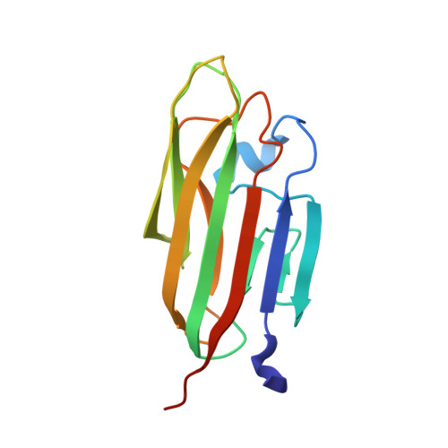
Conformational Plasticity in the Transsynaptic Neurexin-Cerebellin-Glutamate Receptor Adhesion Complex.

Cheng, S.Seven, A.B.Wang, J.Skiniotis, G.Ozkan, E.

(2016) Structure 24: 2163-2173

  • DOI: https://doi.org/10.1016/j.str.2016.11.004
  • Primary Citation of Related Structures:  
    5KWR, 5L2E

  • PubMed Abstract: 

    Synaptic specificity is a defining property of neural networks. In the cerebellum, synapses between parallel fiber neurons and Purkinje cells are specified by the simultaneous interactions of secreted protein cerebellin with pre-synaptic neurexin and post-synaptic delta-type glutamate receptors (GluD). Here, we determined the crystal structures of the trimeric C1q-like domain of rat cerebellin-1, and the first complete ectodomain of a GluD, rat GluD2. Cerebellin binds to the LNS6 domain of α- and β-neurexin-1 through a high-affinity interaction that involves its highly flexible N-terminal domain. In contrast, we show that the interaction of cerebellin with isolated GluD2 ectodomain is low affinity, which is not simply an outcome of lost avidity when compared with binding with a tetrameric full-length receptor. Rather, high-affinity capture of cerebellin by post-synaptic terminals is likely controlled by long-distance regulation within this transsynaptic complex. Altogether, our results suggest unusual conformational flexibility within all components of the complex.


  • Organizational Affiliation

    Department of Biochemistry and Molecular Biology, University of Chicago, Chicago, IL 60637, USA.


Macromolecules
Find similar proteins by:  (by identity cutoff)  |  3D Structure
Entity ID: 1
MoleculeChains Sequence LengthOrganismDetailsImage
Cerebellin-1143Rattus norvegicusMutation(s): 0 
Gene Names: Cbln1
UniProt
Find proteins for P63182 (Rattus norvegicus)
Explore P63182 
Go to UniProtKB:  P63182
Entity Groups  
Sequence Clusters30% Identity50% Identity70% Identity90% Identity95% Identity100% Identity
UniProt GroupP63182
Sequence Annotations
Expand
  • Reference Sequence
Oligosaccharides

Help

Entity ID: 2
MoleculeChains Length2D Diagram Glycosylation3D Interactions
alpha-L-fucopyranose-(1-3)-[2-acetamido-2-deoxy-beta-D-glucopyranose-(1-4)]2-acetamido-2-deoxy-beta-D-glucopyranose
B
3N-Glycosylation
Glycosylation Resources
GlyTouCan:  G11971MR
GlyCosmos:  G11971MR
GlyGen:  G11971MR
Experimental Data & Validation

Experimental Data

Unit Cell:
Length ( Å )Angle ( ˚ )
a = 82.74α = 90
b = 82.74β = 90
c = 50.37γ = 120
Software Package:
Software NamePurpose
PHENIXrefinement
XDSdata reduction
XDSdata scaling
PHASERphasing
Cootmodel building

Structure Validation

View Full Validation Report


Entry History & Funding Information

Deposition Data


Funding OrganizationLocationGrant Number
Klingenstein FoundationUnited States--

Revision History  (Full details and data files)

  • Version 1.0: 2016-08-03
    Type: Initial release
  • Version 1.1: 2016-12-28
    Changes: Database references
  • Version 2.0: 2020-07-29
    Type: Remediation
    Reason: Carbohydrate remediation
    Changes: Atomic model, Data collection, Derived calculations, Structure summary
  • Version 2.1: 2023-10-04
    Changes: Data collection, Database references, Derived calculations, Refinement description, Structure summary