5KDU

ZmpB metallopeptidase in complex with a2,6-Sialyl T-antigen


Experimental Data Snapshot

  • Method: X-RAY DIFFRACTION
  • Resolution: 2.00 Å
  • R-Value Free: 0.201 
  • R-Value Work: 0.169 
  • R-Value Observed: 0.170 

wwPDB Validation   3D Report Full Report


This is version 2.1 of the entry. See complete history


Literature

Recognition of protein-linked glycans as a determinant of peptidase activity.

Noach, I.Ficko-Blean, E.Pluvinage, B.Stuart, C.Jenkins, M.L.Brochu, D.Buenbrazo, N.Wakarchuk, W.Burke, J.E.Gilbert, M.Boraston, A.B.

(2017) Proc Natl Acad Sci U S A 114: E679-E688

  • DOI: https://doi.org/10.1073/pnas.1615141114
  • Primary Citation of Related Structures:  
    5KD2, 5KD5, 5KD8, 5KDJ, 5KDN, 5KDS, 5KDU, 5KDV, 5KDW, 5KDX

  • PubMed Abstract: 

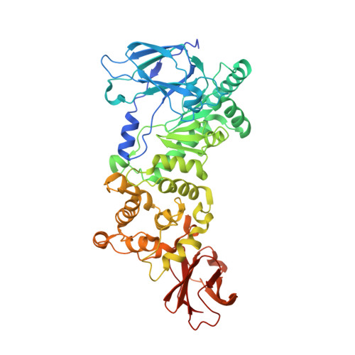
    The vast majority of proteins are posttranslationally altered, with the addition of covalently linked sugars (glycosylation) being one of the most abundant modifications. However, despite the hydrolysis of protein peptide bonds by peptidases being a process essential to all life on Earth, the fundamental details of how peptidases accommodate posttranslational modifications, including glycosylation, has not been addressed. Through biochemical analyses and X-ray crystallographic structures we show that to hydrolyze their substrates, three structurally related metallopeptidases require the specific recognition of O-linked glycan modifications via carbohydrate-specific subsites immediately adjacent to their peptidase catalytic machinery. The three peptidases showed selectivity for different glycans, revealing protein-specific adaptations to particular glycan modifications, yet always cleaved the peptide bond immediately preceding the glycosylated residue. This insight builds upon the paradigm of how peptidases recognize substrates and provides a molecular understanding of glycoprotein degradation.


  • Organizational Affiliation

    Department of Biochemistry and Microbiology, University of Victoria, Victoria, BC V8W 3P6, Canada.


Macromolecules
Find similar proteins by:  (by identity cutoff)  |  3D Structure
Entity ID: 1
MoleculeChains Sequence LengthOrganismDetailsImage
F5/8 type C domain protein530Clostridium perfringens ATCC 13124Mutation(s): 0 
Gene Names: CPF_1489
UniProt
Find proteins for A0A0H2YN38 (Clostridium perfringens (strain ATCC 13124 / DSM 756 / JCM 1290 / NCIMB 6125 / NCTC 8237 / Type A))
Explore A0A0H2YN38 
Go to UniProtKB:  A0A0H2YN38
Entity Groups  
Sequence Clusters30% Identity50% Identity70% Identity90% Identity95% Identity100% Identity
UniProt GroupA0A0H2YN38
Sequence Annotations
Expand
  • Reference Sequence
Oligosaccharides

Help

Entity ID: 2
MoleculeChains Length2D Diagram Glycosylation3D Interactions
beta-D-galactopyranose-(1-3)-[N-acetyl-alpha-neuraminic acid-(2-6)]2-acetamido-2-deoxy-alpha-D-galactopyranose
B
3O-Glycosylation
Glycosylation Resources
GlyTouCan:  G56682BC
GlyCosmos:  G56682BC
GlyGen:  G56682BC
Small Molecules
Ligands 3 Unique
IDChains Name / Formula / InChI Key2D Diagram3D Interactions
SER
Query on SER

Download Ideal Coordinates CCD File 
M [auth A]SERINE
C3 H7 N O3
MTCFGRXMJLQNBG-REOHCLBHSA-N
ZN
Query on ZN

Download Ideal Coordinates CCD File 
L [auth A]ZINC ION
Zn
PTFCDOFLOPIGGS-UHFFFAOYSA-N
EDO
Query on EDO

Download Ideal Coordinates CCD File 
C [auth A]
D [auth A]
E [auth A]
F [auth A]
G [auth A]
C [auth A],
D [auth A],
E [auth A],
F [auth A],
G [auth A],
H [auth A],
I [auth A],
J [auth A],
K [auth A]
1,2-ETHANEDIOL
C2 H6 O2
LYCAIKOWRPUZTN-UHFFFAOYSA-N
Experimental Data & Validation

Experimental Data

  • Method: X-RAY DIFFRACTION
  • Resolution: 2.00 Å
  • R-Value Free: 0.201 
  • R-Value Work: 0.169 
  • R-Value Observed: 0.170 
  • Space Group: P 21 21 21
Unit Cell:
Length ( Å )Angle ( ˚ )
a = 66.14α = 90
b = 68.38β = 90
c = 170.94γ = 90
Software Package:
Software NamePurpose
REFMACrefinement
MOSFLMdata reduction
SCALAdata scaling
MOLREPphasing

Structure Validation

View Full Validation Report


Entry History 

Deposition Data

Revision History  (Full details and data files)

  • Version 1.0: 2017-01-11
    Type: Initial release
  • Version 1.1: 2017-02-01
    Changes: Database references
  • Version 1.2: 2017-02-08
    Changes: Database references
  • Version 2.0: 2020-07-29
    Type: Remediation
    Reason: Carbohydrate remediation
    Changes: Advisory, Atomic model, Data collection, Derived calculations, Non-polymer description, Structure summary
  • Version 2.1: 2023-09-27
    Changes: Data collection, Database references, Refinement description, Structure summary