5KAG

Crystal structure of a dioxygenase in the Crotonase superfamily in P21


Experimental Data Snapshot

  • Method: X-RAY DIFFRACTION
  • Resolution: 2.46 Å
  • R-Value Free: 0.238 
  • R-Value Work: 0.212 
  • R-Value Observed: 0.215 

wwPDB Validation   3D Report Full Report


Ligand Structure Quality Assessment 


This is version 1.3 of the entry. See complete history

Re-refinement Note

This entry reflects an alternative modeling of the original data in: 2NP9


Literature

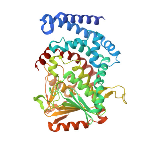
Probing the structural basis of oxygen binding in a cofactor-independent dioxygenase.

Li, K.Fielding, E.N.Condurso, H.L.Bruner, S.D.

(2017) Acta Crystallogr D Struct Biol 73: 573-580

  • DOI: https://doi.org/10.1107/S2059798317007045
  • Primary Citation of Related Structures:  
    5KAG, 5KAH, 5KAJ

  • PubMed Abstract: 

    The enzyme DpgC is included in the small family of cofactor-independent dioxygenases. The chemistry of DpgC is uncommon as the protein binds and utilizes dioxygen without the aid of a metal or organic cofactor. Previous structural and biochemical studies identified the substrate-binding mode and the components of the active site that are important in the catalytic mechanism. In addition, the results delineated a putative binding pocket and migration pathway for the co-substrate dioxygen. Here, structural biology is utilized, along with site-directed mutagenesis, to probe the assigned dioxygen-binding pocket. The key residues implicated in dioxygen trafficking were studied to probe the process of binding, activation and chemistry. The results support the proposed chemistry and provide insight into the general mechanism of dioxygen binding and activation.


  • Organizational Affiliation

    Department of Chemistry, University of Florida, Gainesville, FL 32611, USA.


Macromolecules
Find similar proteins by:  (by identity cutoff)  |  3D Structure
Entity ID: 1
MoleculeChains Sequence LengthOrganismDetailsImage
(3,5-dihydroxyphenyl)acetyl-CoA 1,2-dioxygenase
A, B, C, D, E
A, B, C, D, E, F, G, H, I, J, K, L
438Streptomyces toyocaensisMutation(s): 0 
Gene Names: BU52_01220
EC: 1.13.11.80
UniProt
Find proteins for Q8KLK7 (Streptomyces toyocaensis)
Explore Q8KLK7 
Go to UniProtKB:  Q8KLK7
Entity Groups  
Sequence Clusters30% Identity50% Identity70% Identity90% Identity95% Identity100% Identity
UniProt GroupQ8KLK7
Sequence Annotations
Expand
  • Reference Sequence
Small Molecules
Ligands 2 Unique
IDChains Name / Formula / InChI Key2D Diagram3D Interactions
YE1
Query on YE1

Download Ideal Coordinates CCD File 
AA [auth H]
CA [auth I]
EA [auth J]
GA [auth K]
IA [auth L]
AA [auth H],
CA [auth I],
EA [auth J],
GA [auth K],
IA [auth L],
M [auth A],
O [auth B],
Q [auth C],
S [auth D],
U [auth E],
W [auth F],
Y [auth G]
[(2R,3S,4R,5R)-5-(6-AMINO-9H-PURIN-9-YL)-4-HYDROXY-3-(PHOSPHONOOXY)TETRAHYDROFURAN-2-YL]METHYL (3R)-4-({3-[(2-{[(3,5-DIHYDROXYPHENYL)ACETYL]AMINO}ETHYL)AMINO]-3-OXOPROPYL}AMINO)-3-HYDROXY-2,2-DIMETHYL-4-OXOBUTYL DIHYDROGEN DIPHOSPHATE
C29 H43 N8 O19 P3
MSJGSRHUBFYIJV-CECATXLMSA-N
OXY
Query on OXY

Download Ideal Coordinates CCD File 
BA [auth H]
DA [auth I]
FA [auth J]
HA [auth K]
JA [auth L]
BA [auth H],
DA [auth I],
FA [auth J],
HA [auth K],
JA [auth L],
N [auth A],
P [auth B],
R [auth C],
T [auth D],
V [auth E],
X [auth F],
Z [auth G]
OXYGEN MOLECULE
O2
MYMOFIZGZYHOMD-UHFFFAOYSA-N
Experimental Data & Validation

Experimental Data

  • Method: X-RAY DIFFRACTION
  • Resolution: 2.46 Å
  • R-Value Free: 0.238 
  • R-Value Work: 0.212 
  • R-Value Observed: 0.215 
  • Space Group: P 1 21 1
Unit Cell:
Length ( Å )Angle ( ˚ )
a = 138.959α = 90
b = 170.447β = 90.06
c = 156.283γ = 90
Software Package:
Software NamePurpose
PHENIXrefinement
XDSdata reduction
Aimlessdata scaling
PHASERphasing

Structure Validation

View Full Validation Report


Ligand Structure Quality Assessment 


Entry History & Funding Information

Deposition Data


Funding OrganizationLocationGrant Number
National Institutes of Health/National Institute of General Medical Sciences (NIH/NIGMS)United StatesGM086570

Revision History  (Full details and data files)

  • Version 1.0: 2017-06-21
    Type: Initial release
  • Version 1.1: 2017-07-26
    Changes: Author supporting evidence, Database references
  • Version 1.2: 2019-12-25
    Changes: Author supporting evidence
  • Version 1.3: 2023-09-27
    Changes: Data collection, Database references, Refinement description