5K8K

Structure of the Haemophilus influenzae LpxH-lipid X complex


Experimental Data Snapshot

  • Method: X-RAY DIFFRACTION
  • Resolution: 2.55 Å
  • R-Value Free: 0.227 
  • R-Value Work: 0.192 
  • R-Value Observed: 0.195 

wwPDB Validation   3D Report Full Report


Ligand Structure Quality Assessment 


This is version 1.4 of the entry. See complete history


Literature

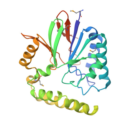
Structure of the essential Haemophilus influenzae UDP-diacylglucosamine pyrophosphohydrolase LpxH in lipid A biosynthesis.

Cho, J.Lee, C.J.Zhao, J.Young, H.E.Zhou, P.

(2016) Nat Microbiol 1: 16154-16154

  • DOI: https://doi.org/10.1038/nmicrobiol.2016.154
  • Primary Citation of Related Structures:  
    5K8K

  • PubMed Abstract: 

    In most Gram-negative pathogens, the hydrolysis of UDP-2,3-diacylglucosamine to generate lipid X in lipid A biosynthesis is catalysed by the membrane-associated enzyme LpxH. We report the crystal structure of LpxH in complex with its product, lipid X, unveiling a unique insertion lid above the conserved architecture of calcineurin-like phosphoesterases. This structure reveals elaborate interactions surrounding lipid X and provides molecular insights into the substrate selectivity, catalysis and inhibition of LpxH.


  • Organizational Affiliation

    Department of Biochemistry, Duke University Medical Center, Research Drive, Durham, North Carolina 27710, USA.


Macromolecules
Find similar proteins by:  (by identity cutoff)  |  3D Structure
Entity ID: 1
MoleculeChains Sequence LengthOrganismDetailsImage
UDP-2,3-diacylglucosamine hydrolase256Haemophilus influenzae Rd KW20Mutation(s): 0 
Gene Names: lpxHHI_0735
EC: 3.6.1.54
Membrane Entity: Yes 
UniProt
Find proteins for P44046 (Haemophilus influenzae (strain ATCC 51907 / DSM 11121 / KW20 / Rd))
Explore P44046 
Go to UniProtKB:  P44046
Entity Groups  
Sequence Clusters30% Identity50% Identity70% Identity90% Identity95% Identity100% Identity
UniProt GroupP44046
Sequence Annotations
Expand
  • Reference Sequence
Small Molecules
Ligands 4 Unique
IDChains Name / Formula / InChI Key2D Diagram3D Interactions
LP5
Query on LP5

Download Ideal Coordinates CCD File 
D [auth A](R)-((2R,3S,4R,5R,6R)-3-HYDROXY-2-(HYDROXYMETHYL)-5-((R)-3-HYDROXYTETRADECANAMIDO)-6-(PHOSPHONOOXY)TETRAHYDRO-2H-PYRAN-4-YL) 3-HYDROXYTETRADECANOATE
C34 H66 N O12 P
HEHQDWUWJVPREQ-XQJZMFRCSA-N
GOL
Query on GOL

Download Ideal Coordinates CCD File 
G [auth A],
H [auth A]
GLYCEROL
C3 H8 O3
PEDCQBHIVMGVHV-UHFFFAOYSA-N
ACT
Query on ACT

Download Ideal Coordinates CCD File 
E [auth A],
F [auth A]
ACETATE ION
C2 H3 O2
QTBSBXVTEAMEQO-UHFFFAOYSA-M
MN
Query on MN

Download Ideal Coordinates CCD File 
B [auth A],
C [auth A]
MANGANESE (II) ION
Mn
WAEMQWOKJMHJLA-UHFFFAOYSA-N
Modified Residues  1 Unique
IDChains TypeFormula2D DiagramParent
MSE
Query on MSE
A
L-PEPTIDE LINKINGC5 H11 N O2 SeMET
Experimental Data & Validation

Experimental Data

  • Method: X-RAY DIFFRACTION
  • Resolution: 2.55 Å
  • R-Value Free: 0.227 
  • R-Value Work: 0.192 
  • R-Value Observed: 0.195 
  • Space Group: H 3 2
Unit Cell:
Length ( Å )Angle ( ˚ )
a = 99.84α = 90
b = 99.84β = 90
c = 200.85γ = 120
Software Package:
Software NamePurpose
PHENIXrefinement
Aimlessdata scaling
XSCALEdata scaling
PDB_EXTRACTdata extraction
XDSdata reduction
PHENIXphasing

Structure Validation

View Full Validation Report


Ligand Structure Quality Assessment 


Entry History & Funding Information

Deposition Data


Funding OrganizationLocationGrant Number
National Institutes of Health/National Institute of General Medical Sciences (NIH/NIGMS)United StatesGM115355
National Institutes of Health/National Institute of General Medical Sciences (NIH/NIGMS)United StatesGM51310

Revision History  (Full details and data files)

  • Version 1.0: 2016-08-10
    Type: Initial release
  • Version 1.1: 2016-09-21
    Changes: Database references
  • Version 1.2: 2016-11-09
    Changes: Database references
  • Version 1.3: 2017-09-20
    Changes: Author supporting evidence, Derived calculations
  • Version 1.4: 2019-12-25
    Changes: Author supporting evidence