5HQG

WD40 domain of Human E3 Ubiquitin Ligase COP1 (RFWD2)


Experimental Data Snapshot

  • Method: X-RAY DIFFRACTION
  • Resolution: 2.00 Å
  • R-Value Free: 0.242 
  • R-Value Work: 0.210 
  • R-Value Observed: 0.214 

wwPDB Validation   3D Report Full Report


This is version 1.4 of the entry. See complete history


Literature

Structural Basis for Substrate Selectivity of the E3 Ligase COP1.

Uljon, S.Xu, X.Durzynska, I.Stein, S.Adelmant, G.Marto, J.A.Pear, W.S.Blacklow, S.C.

(2016) Structure 24: 687-696

  • DOI: https://doi.org/10.1016/j.str.2016.03.002
  • Primary Citation of Related Structures:  
    5HQG, 5IGO, 5IGQ

  • PubMed Abstract: 

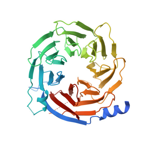
    COP1 proteins are E3 ubiquitin ligases that regulate phototropism in plants and target transcription factors for degradation in mammals. The substrate-binding region of COP1 resides within a WD40-repeat domain that also binds to Trib proteins, which are adaptors for C/EBPα degradation. Here we report structures of the human COP1 WD40 domain in isolation, and complexes of the human and Arabidopsis thaliana COP1 WD40 domains with the binding motif of Trib1. The human and Arabidopsis WD40 domains are seven-bladed β propellers with an inserted loop on the bottom face of the first blade. The Trib1 peptide binds in an extended conformation to a highly conserved surface on the top face of the β propeller, indicating a general mode for recognition of peptide motifs by COP1. Together, these studies identify the structural basis and key interactions for motif recognition by COP1, and hint at how Trib1 autoinhibition is overcome to target C/EBPα for degradation.


  • Organizational Affiliation

    Department of Cancer Biology, Dana Farber Cancer Institute, Boston, MA 02215, USA; Department of Biological Chemistry and Molecular Pharmacology, Harvard Medical School, Boston, MA 02115, USA; Department of Pathology, Brigham and Women's Hospital and Harvard Medical School, Boston, MA 02115, USA.


Macromolecules
Find similar proteins by:  (by identity cutoff)  |  3D Structure
Entity ID: 1
MoleculeChains Sequence LengthOrganismDetailsImage
E3 ubiquitin-protein ligase RFWD2362Homo sapiensMutation(s): 0 
Gene Names: RFWD2COP1RNF200
EC: 6.3.2
UniProt & NIH Common Fund Data Resources
Find proteins for Q8NHY2 (Homo sapiens)
Explore Q8NHY2 
Go to UniProtKB:  Q8NHY2
PHAROS:  Q8NHY2
GTEx:  ENSG00000143207 
Entity Groups  
Sequence Clusters30% Identity50% Identity70% Identity90% Identity95% Identity100% Identity
UniProt GroupQ8NHY2
Sequence Annotations
Expand
  • Reference Sequence
Experimental Data & Validation

Experimental Data

Unit Cell:
Length ( Å )Angle ( ˚ )
a = 68.914α = 90
b = 68.914β = 90
c = 133.922γ = 120
Software Package:
Software NamePurpose
PHENIXrefinement
XDSdata reduction
Aimlessdata scaling
PHASERphasing

Structure Validation

View Full Validation Report


Entry History & Funding Information

Deposition Data


Funding OrganizationLocationGrant Number
National Institutes of Health/National Cancer Institute (NIH/NCI)United StatesK08 CA166227

Revision History  (Full details and data files)

  • Version 1.0: 2016-04-20
    Type: Initial release
  • Version 1.1: 2016-05-11
    Changes: Database references
  • Version 1.2: 2017-09-20
    Changes: Author supporting evidence, Database references, Derived calculations
  • Version 1.3: 2019-12-04
    Changes: Author supporting evidence
  • Version 1.4: 2023-09-27
    Changes: Data collection, Database references, Refinement description