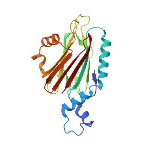
A novel sphingomyelin/cholesterol domain-specific probe reveals the dynamics of the membrane domains during virus release and in Niemann-Pick type C
Makino, A., Abe, M., Ishitsuka, R., Murate, M., Kishimoto, T., Sakai, S., Hullin-Matsuda, F., Shimada, Y., Inaba, T., Miyatake, H., Tanaka, H., Kurahashi, A., Pack, C.G., Kasai, R.S., Kubo, S., Schieber, N.L., Dohmae, N., Tochio, N., Hagiwara, K., Sasaki, Y., Aida, Y., Fujimori, F., Kigawa, T., Nishibori, K., Parton, R.G., Kusumi, A., Sako, Y., Anderluh, G., Yamashita, M., Kobayashi, T., Greimel, P., Kobayashi, T.(2017) FASEB J 31: 1301-1322
- PubMed: 27492925
- DOI: https://doi.org/10.1096/fj.201500075R
- Primary Citation of Related Structures:
5H0Q - PubMed Abstract:
We identified a novel, nontoxic mushroom protein that specifically binds to a complex of sphingomyelin (SM), a major sphingolipid in mammalian cells, and cholesterol (Chol). The purified protein, termed nakanori, labeled cell surface domains in an SM- and Chol-dependent manner and decorated specific lipid domains that colocalized with inner leaflet small GTPase H-Ras, but not K-Ras. The use of nakanori as a lipid-domain-specific probe revealed altered distribution and dynamics of SM/Chol on the cell surface of Niemann-Pick type C fibroblasts, possibly explaining some of the disease phenotype. In addition, that nakanori treatment of epithelial cells after influenza virus infection potently inhibited virus release demonstrates the therapeutic value of targeting specific lipid domains for anti-viral treatment.-Makino, A., Abe, M., Ishitsuka, R., Murate, M., Kishimoto, T., Sakai, S., Hullin-Matsuda, F., Shimada, Y., Inaba, T., Miyatake, H., Tanaka, H., Kurahashi, A., Pack, C.-G., Kasai, R. S., Kubo, S., Schieber, N. L., Dohmae, N., Tochio, N., Hagiwara, K., Sasaki, Y., Aida, Y., Fujimori, F., Kigawa, T., Nishibori, K., Parton, R. G., Kusumi, A., Sako, Y., Anderluh, G., Yamashita, M., Kobayashi, T., Greimel, P., Kobayashi, T. A novel sphingomyelin/cholesterol domain-specific probe reveals the dynamics of the membrane domains during virus release and in Niemann-Pick type C.
Organizational Affiliation:
Rikagaku Kenkyūsho (RIKEN), Saitama, Japan.


