5EIA

mACHE-anti TZ2PA5 complex from a 1:6 mixture of the syn/anti isomers


Experimental Data Snapshot

  • Method: X-RAY DIFFRACTION
  • Resolution: 2.70 Å
  • R-Value Free: 0.208 
  • R-Value Work: 0.176 
  • R-Value Observed: 0.176 

wwPDB Validation   3D Report Full Report


Ligand Structure Quality Assessment 


This is version 1.3 of the entry. See complete history


Literature

Steric and Dynamic Parameters Influencing In Situ Cycloadditions to Form Triazole Inhibitors with Crystalline Acetylcholinesterase.

Bourne, Y.Sharpless, K.B.Taylor, P.Marchot, P.

(2016) J Am Chem Soc 138: 1611-1621

  • DOI: https://doi.org/10.1021/jacs.5b11384
  • Primary Citation of Related Structures:  
    5EHN, 5EHQ, 5EHZ, 5EIA, 5EIE, 5EIH

  • PubMed Abstract: 

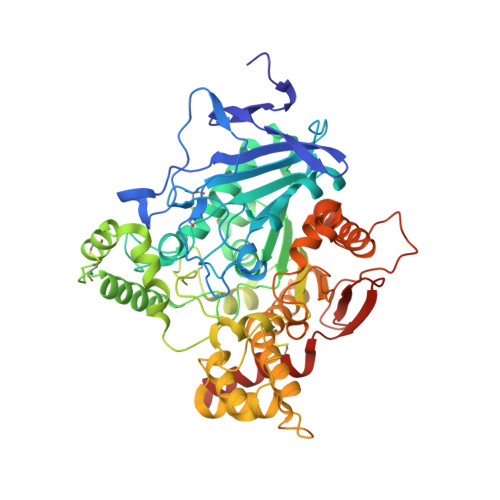
    Ligand binding sites on acetylcholinesterase (AChE) comprise an active center, at the base of a deep and narrow gorge lined by aromatic residues, and a peripheral site at the gorge entry. These features launched AChE as a reaction vessel for in situ click-chemistry synthesis of high-affinity TZ2PA6 and TZ2PA5 inhibitors, forming a syn-triazole upon cycloaddition within the gorge from alkyne and azide reactants bound at the two sites, respectively. Subsequent crystallographic analyses of AChE complexes with the TZ2PA6 regioisomers demonstrated that syn product association is accompanied by side chain reorganization within the gorge, freezing-in-frame a conformation distinct from an unbound state or anti complex. To correlate inhibitor dimensions with reactivity and explore whether in situ cycloaddition could be accelerated in a concentrated, crystalline template, we developed crystal-soaking procedures and solved structures of AChE complexes with the TZ2PA5 regioisomers and their TZ2/PA5 precursors (2.1-2.7 Å resolution). The structures reveal motions of residue His447 in the active site and, unprecedentedly, residue Tyr341 at the gorge mouth, associated with TZ2 binding and coordinated with other side chain motions in the gorge that may guide AChE toward a transient state favoring syn-triazole formation. Despite precursor binding to crystalline AChE, coupling of rapid electric field fluctuations in the gorge with proper alignments of the azide and alkyne reactants to form the triazole remains a likely limiting step. These observations point to a prime requirement for AChE to interconvert dynamically between sequential conformations to promote favorable electrostatic factors enabling a productive apposition of the reactants for reactivity.


  • Organizational Affiliation

    Aix-Marseille Université, laboratory Architecture et Fonction des Macromolécules Biologiques, Faculté des Sciences de Luminy , 13288 Marseille cedex 09, France.


Macromolecules
Find similar proteins by:  (by identity cutoff)  |  3D Structure
Entity ID: 1
MoleculeChains Sequence LengthOrganismDetailsImage
Acetylcholinesterase
A, B
543Mus musculusMutation(s): 0 
Gene Names: Ache
EC: 3.1.1.7
UniProt & NIH Common Fund Data Resources
Find proteins for P21836 (Mus musculus)
Explore P21836 
Go to UniProtKB:  P21836
IMPC:  MGI:87876
Entity Groups  
Sequence Clusters30% Identity50% Identity70% Identity90% Identity95% Identity100% Identity
UniProt GroupP21836
Sequence Annotations
Expand
  • Reference Sequence
Small Molecules
Ligands 3 Unique
IDChains Name / Formula / InChI Key2D Diagram3D Interactions
5O2
Query on 5O2

Download Ideal Coordinates CCD File 
E [auth A],
I [auth B]
6-phenyl-5-[5-[1-[2-(1,2,3,4-tetrahydroacridin-9-ylamino)ethyl]-1,2,3-triazol-4-yl]pentyl]phenanthridin-5-ium-3,8-diamine
C41 H43 N8
XIORYVGXAQRYQA-UHFFFAOYSA-O
NAG
Query on NAG

Download Ideal Coordinates CCD File 
C [auth A],
D [auth A],
G [auth B],
H [auth B]
2-acetamido-2-deoxy-beta-D-glucopyranose
C8 H15 N O6
OVRNDRQMDRJTHS-FMDGEEDCSA-N
PG4
Query on PG4

Download Ideal Coordinates CCD File 
F [auth A]TETRAETHYLENE GLYCOL
C8 H18 O5
UWHCKJMYHZGTIT-UHFFFAOYSA-N
Experimental Data & Validation

Experimental Data

  • Method: X-RAY DIFFRACTION
  • Resolution: 2.70 Å
  • R-Value Free: 0.208 
  • R-Value Work: 0.176 
  • R-Value Observed: 0.176 
  • Space Group: P 21 21 21
Unit Cell:
Length ( Å )Angle ( ˚ )
a = 79.47α = 90
b = 112.52β = 90
c = 226.56γ = 90
Software Package:
Software NamePurpose
BUSTERrefinement
XDSdata reduction
SCALAdata scaling
REFMACphasing

Structure Validation

View Full Validation Report


Ligand Structure Quality Assessment 


Entry History 

Deposition Data

Revision History  (Full details and data files)

  • Version 1.0: 2016-01-20
    Type: Initial release
  • Version 1.1: 2016-02-17
    Changes: Database references
  • Version 1.2: 2020-07-29
    Type: Remediation
    Reason: Carbohydrate remediation
    Changes: Data collection, Derived calculations, Structure summary
  • Version 1.3: 2024-01-10
    Changes: Data collection, Database references, Refinement description, Structure summary