5D2A

Bifunctional dendrimers


Experimental Data Snapshot

  • Method: X-RAY DIFFRACTION
  • Resolution: 2.13 Å
  • R-Value Free: 0.246 
  • R-Value Work: 0.205 
  • R-Value Observed: 0.209 

wwPDB Validation   3D Report Full Report


Ligand Structure Quality Assessment 


This is version 1.4 of the entry. See complete history


Literature

Overcoming antibiotic resistance inPseudomonas aeruginosabiofilms using glycopeptide dendrimers.

Michaud, G.Visini, R.Bergmann, M.Salerno, G.Bosco, R.Gillon, E.Richichi, B.Nativi, C.Imberty, A.Stocker, A.Darbre, T.Reymond, J.L.

(2016) Chem Sci 7: 166-182

  • DOI: https://doi.org/10.1039/c5sc03635f
  • Primary Citation of Related Structures:  
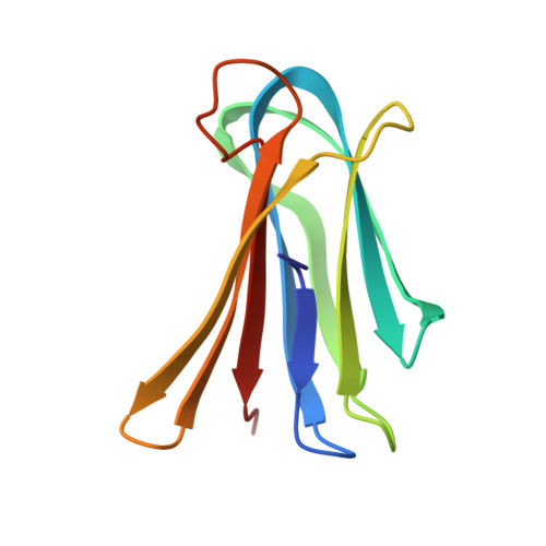
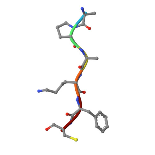
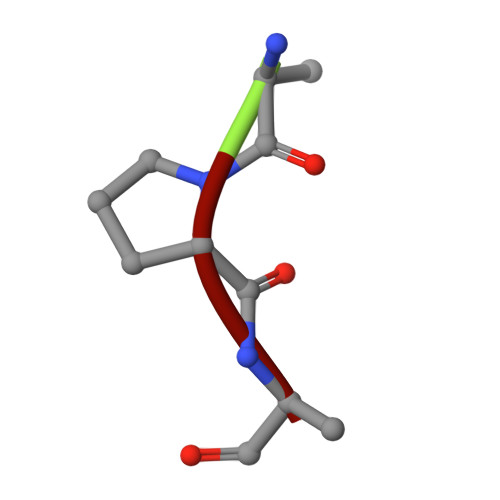
    5D2A

  • PubMed Abstract: 

    Antibiotic resistance in the opportunistic pathogen Pseudomonas aeruginosa is partly caused by biofilms forming a physical barrier to antibiotic penetration. Here we focused on modifying tetravalent glycopeptide dendrimer ligands of P. aeruginosa lectins LecB or LecA to increase their biofilm inhibition activity. First heteroglycoclusters were investigated displaying one pair each of LecB specific fucosyl groups and LecA specific galactosyl groups and binding simultaneously to both lectins, one of which gave the first fully resolved crystal structure of a peptide dendrimer as LecB complex providing a structural model for dendrimer-lectin interactions (PDB ; 5D2A). Biofilm inhibition was increased by introducing additional cationic residues in these dendrimers but resulted in bactericidal effects similar to those of non-glycosylated polycationic antimicrobial peptide dendrimers. In a second approach dendrimers displaying four copies of the natural LecB ligand Lewis a were prepared leading to slightly stronger LecB binding and biofilm inhibition. Finally synergistic application of a LecB specific non-bactericidal antibiofilm dendrimer with the antibiotic tobramycin at sub-inhibitory concentrations of both compounds allowed effective biofilm inhibition and dispersal.


  • Organizational Affiliation

    Department of Chemistry and Biochemistry , University of Berne , Freiestrasse 3 , 3012 Berne , Switzerland . Email: jean-louis.reymond@dcb.unibe.ch.


Macromolecules
Find similar proteins by:  (by identity cutoff)  |  3D Structure
Entity ID: 1
MoleculeChains Sequence LengthOrganismDetailsImage
Fucose-binding lectin
A, B
114Pseudomonas aeruginosaMutation(s): 0 
Gene Names: lecBERS445055_01627PA8380_17510PAE221_03716PAMH19_1713YQ19_10010
UniProt
Find proteins for Q9HYN5 (Pseudomonas aeruginosa (strain ATCC 15692 / DSM 22644 / CIP 104116 / JCM 14847 / LMG 12228 / 1C / PRS 101 / PAO1))
Explore Q9HYN5 
Go to UniProtKB:  Q9HYN5
Entity Groups  
Sequence Clusters30% Identity50% Identity70% Identity90% Identity95% Identity100% Identity
UniProt GroupQ9HYN5
Sequence Annotations
Expand
  • Reference Sequence

Find similar proteins by:  Sequence   |   3D Structure  

Entity ID: 2
MoleculeChains Sequence LengthOrganismDetailsImage
ZDC-ALA-PRO-ALA-LYS-PHE-CYS-ALA-PRO-ALA-PHB-GAL6synthetic constructMutation(s): 0 
Sequence Annotations
Expand
  • Reference Sequence

Find similar proteins by:  Sequence   |   3D Structure  

Entity ID: 3
MoleculeChains Sequence LengthOrganismDetailsImage
ZDC-ALA-PRO-ALA-LYS-PHE-CYS-ALA-PRO-ALA-PHB-GAL
D, E
3synthetic constructMutation(s): 0 
Sequence Annotations
Expand
  • Reference Sequence
Small Molecules
Ligands 4 Unique
IDChains Name / Formula / InChI Key2D Diagram3D Interactions
ZDC
Query on ZDC

Download Ideal Coordinates CCD File 
L [auth D],
M [auth E]
3,7-anhydro-2,8-dideoxy-L-glycero-D-gluco-octonic acid
C8 H14 O6
YTZUDUWVQZSKNN-OASCRQMUSA-N
GAL
Query on GAL

Download Ideal Coordinates CCD File 
K [auth C]beta-D-galactopyranose
C6 H12 O6
WQZGKKKJIJFFOK-FPRJBGLDSA-N
PHB
Query on PHB

Download Ideal Coordinates CCD File 
J [auth C]P-HYDROXYBENZOIC ACID
C7 H6 O3
FJKROLUGYXJWQN-UHFFFAOYSA-N
CA
Query on CA

Download Ideal Coordinates CCD File 
F [auth A],
G [auth A],
H [auth B],
I [auth B]
CALCIUM ION
Ca
BHPQYMZQTOCNFJ-UHFFFAOYSA-N
Experimental Data & Validation

Experimental Data

  • Method: X-RAY DIFFRACTION
  • Resolution: 2.13 Å
  • R-Value Free: 0.246 
  • R-Value Work: 0.205 
  • R-Value Observed: 0.209 
  • Space Group: P 63 2 2
Unit Cell:
Length ( Å )Angle ( ˚ )
a = 105.107α = 90
b = 105.107β = 90
c = 111.092γ = 120
Software Package:
Software NamePurpose
PHASERphasing
PHENIXrefinement
PDB_EXTRACTdata extraction
XDSdata reduction
XDSdata scaling

Structure Validation

View Full Validation Report


Ligand Structure Quality Assessment 


Entry History 

Deposition Data

Revision History  (Full details and data files)

  • Version 1.0: 2016-02-10
    Type: Initial release
  • Version 1.1: 2018-07-11
    Changes: Data collection, Database references
  • Version 1.2: 2019-05-08
    Changes: Advisory, Data collection, Derived calculations
  • Version 1.3: 2020-07-29
    Type: Remediation
    Reason: Carbohydrate remediation
    Changes: Data collection, Derived calculations, Structure summary
  • Version 1.4: 2024-01-10
    Changes: Data collection, Database references, Refinement description, Structure summary