5CYU

Structure of the soluble domain of EccB1 from the Mycobacterium smegmatis ESX-1 secretion system.


Experimental Data Snapshot

  • Method: X-RAY DIFFRACTION
  • Resolution: 3.07 Å
  • R-Value Free: 0.297 
  • R-Value Work: 0.241 
  • R-Value Observed: 0.246 

wwPDB Validation   3D Report Full Report


This is version 1.6 of the entry. See complete history


Literature

Structures of EccB1 and EccD1 from the core complex of the mycobacterial ESX-1 type VII secretion system.

Wagner, J.M.Chan, S.Evans, T.J.Kahng, S.Kim, J.Arbing, M.A.Eisenberg, D.Korotkov, K.V.

(2016) BMC Struct Biol 16: 5-5

  • DOI: https://doi.org/10.1186/s12900-016-0056-6
  • Primary Citation of Related Structures:  
    4KK7, 4KV2, 4KV3, 5CYU

  • PubMed Abstract: 

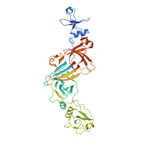
    The ESX-1 type VII secretion system is an important determinant of virulence in pathogenic mycobacteria, including Mycobacterium tuberculosis. This complicated molecular machine secretes folded proteins through the mycobacterial cell envelope to subvert the host immune response. Despite its important role in disease very little is known about the molecular architecture of the ESX-1 secretion system. This study characterizes the structures of the soluble domains of two conserved core ESX-1 components - EccB1 and EccD1. The periplasmic domain of EccB1 consists of 4 repeat domains and a central domain, which together form a quasi 2-fold symmetrical structure. The repeat domains of EccB1 are structurally similar to a known peptidoglycan binding protein suggesting a role in anchoring the ESX-1 system within the periplasmic space. The cytoplasmic domain of EccD1has a ubiquitin-like fold and forms a dimer with a negatively charged groove. These structures represent a major step towards resolving the molecular architecture of the entire ESX-1 assembly and may contribute to ESX-1 targeted tuberculosis intervention strategies.


  • Organizational Affiliation

    Department of Molecular & Cellular Biochemistry and Center for Structural Biology, University of Kentucky, 741 South Limestone, Lexington, KY, 40536, USA. jonw7782@gmail.com.


Macromolecules
Find similar proteins by:  (by identity cutoff)  |  3D Structure
Entity ID: 1
MoleculeChains Sequence LengthOrganismDetailsImage
Conserved membrane protein408Mycolicibacterium smegmatis MC2 155Mutation(s): 0 
Gene Names: MSMEG_0060MSMEI_0061
UniProt
Find proteins for A0QNJ0 (Mycolicibacterium smegmatis (strain ATCC 700084 / mc(2)155))
Explore A0QNJ0 
Go to UniProtKB:  A0QNJ0
Entity Groups  
Sequence Clusters30% Identity50% Identity70% Identity90% Identity95% Identity100% Identity
UniProt GroupA0QNJ0
Sequence Annotations
Expand
  • Reference Sequence
Experimental Data & Validation

Experimental Data

  • Method: X-RAY DIFFRACTION
  • Resolution: 3.07 Å
  • R-Value Free: 0.297 
  • R-Value Work: 0.241 
  • R-Value Observed: 0.246 
  • Space Group: P 61 2 2
Unit Cell:
Length ( Å )Angle ( ˚ )
a = 74.41α = 90
b = 74.41β = 90
c = 280.6γ = 120
Software Package:
Software NamePurpose
XDSdata reduction
XSCALEdata scaling
PHASERphasing
Cootmodel building
BUSTER-TNTrefinement
PDB_EXTRACTdata extraction

Structure Validation

View Full Validation Report


Entry History & Funding Information

Deposition Data


Funding OrganizationLocationGrant Number
National Institutes of Health/National Institute Of Allergy and Infectious Diseases (NIH/NIAID)United StatesTBSGC R01 (A1068135)
National Institutes of Health/National Institute Of Allergy and Infectious Diseases (NIH/NIAID)United StatesTBSGC P01 (AI095208)

Revision History  (Full details and data files)

  • Version 1.0: 2015-08-12
    Type: Initial release
  • Version 1.1: 2016-03-23
    Changes: Database references
  • Version 1.2: 2016-07-27
    Changes: Data collection
  • Version 1.3: 2017-09-13
    Changes: Author supporting evidence, Derived calculations
  • Version 1.4: 2019-12-11
    Changes: Author supporting evidence
  • Version 1.5: 2022-03-30
    Changes: Author supporting evidence, Database references
  • Version 1.6: 2023-09-27
    Changes: Data collection, Refinement description