5C0Y

Crystal structure of the Rrp6 catalytic domain bound to poly(U) RNA


Experimental Data Snapshot

  • Method: X-RAY DIFFRACTION
  • Resolution: 2.10 Å
  • R-Value Free: 0.180 
  • R-Value Work: 0.148 
  • R-Value Observed: 0.151 

wwPDB Validation   3D Report Full Report


This is version 1.2 of the entry. See complete history


Literature

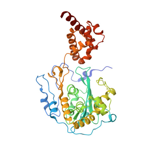
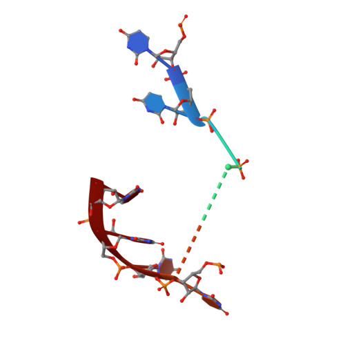
RNA degradation paths in a 12-subunit nuclear exosome complex.

Makino, D.L.Schuch, B.Stegmann, E.Baumgartner, M.Basquin, C.Conti, E.

(2015) Nature 524: 54-58

  • DOI: https://doi.org/10.1038/nature14865
  • Primary Citation of Related Structures:  
    5C0W, 5C0X, 5C0Y

  • PubMed Abstract: 

    The eukaryotic exosome is a conserved RNA-degrading complex that functions in RNA surveillance, turnover and processing. How the same machinery can either completely degrade or precisely trim RNA substrates has long remained unexplained. Here we report the crystal structures of a yeast nuclear exosome containing the 9-subunit core, the 3'-5' RNases Rrp44 and Rrp6, and the obligate Rrp6-binding partner Rrp47 in complex with different RNAs. The combined structural and biochemical data of this 12-subunit complex reveal how a single-stranded RNA can reach the Rrp44 or Rrp6 active sites directly or can bind Rrp6 and be threaded via the central channel towards the distal RNase Rrp44. When a bulky RNA is stalled at the entrance of the channel, Rrp6-Rrp47 swings open. The results suggest how the same molecular machine can coordinate processive degradation and partial trimming in an RNA-dependent manner by a concerted swinging mechanism of the two RNase subunits.


  • Organizational Affiliation

    Department of Structural Cell Biology, Max Planck Institute of Biochemistry, 82152 Martinsried, Germany.


Macromolecules

Find similar proteins by:  (by identity cutoff)  |  3D Structure
Entity ID: 1
MoleculeChains Sequence LengthOrganismDetailsImage
Exosome complex exonuclease RRP6
A, B
402Saccharomyces cerevisiae S288CMutation(s): 1 
Gene Names: RRP6UNC733YOR001W
EC: 3.1.13
UniProt
Find proteins for Q12149 (Saccharomyces cerevisiae (strain ATCC 204508 / S288c))
Explore Q12149 
Go to UniProtKB:  Q12149
Entity Groups  
Sequence Clusters30% Identity50% Identity70% Identity90% Identity95% Identity100% Identity
UniProt GroupQ12149
Sequence Annotations
Expand
  • Reference Sequence

Find similar nucleic acids by:  Sequence   |   3D Structure  

Entity ID: 2
MoleculeChains LengthOrganismImage
poly U RNAC [auth D],
D [auth C]
15synthetic construct
Sequence Annotations
Expand
  • Reference Sequence
Small Molecules
Ligands 1 Unique
IDChains Name / Formula / InChI Key2D Diagram3D Interactions
MG
Query on MG

Download Ideal Coordinates CCD File 
E [auth B]MAGNESIUM ION
Mg
JLVVSXFLKOJNIY-UHFFFAOYSA-N
Experimental Data & Validation

Experimental Data

  • Method: X-RAY DIFFRACTION
  • Resolution: 2.10 Å
  • R-Value Free: 0.180 
  • R-Value Work: 0.148 
  • R-Value Observed: 0.151 
  • Space Group: P 31
Unit Cell:
Length ( Å )Angle ( ˚ )
a = 110.186α = 90
b = 110.186β = 90
c = 78.883γ = 120
Software Package:
Software NamePurpose
XDSdata reduction
Aimlessdata scaling
PHASERphasing
PHENIXrefinement
Cootmodel building
PDB_EXTRACTdata extraction

Structure Validation

View Full Validation Report


Entry History & Funding Information

Deposition Data


Funding OrganizationLocationGrant Number
European Research CouncilGermanyAdvanced Investigator Grant 294371
Marie CurieGermanyITN RNPnet
German Research FoundationGermanySFB646, SFB1035, GRK1721, FOR1680 and CIPSM
Louis Jeantet FoundationSwitzerland--

Revision History  (Full details and data files)

  • Version 1.0: 2015-08-05
    Type: Initial release
  • Version 1.1: 2015-08-12
    Changes: Database references
  • Version 1.2: 2017-09-06
    Changes: Author supporting evidence