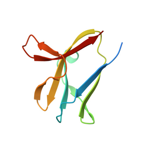
4YEF

beta1 carbohydrate binding module (CBM) of AMP-activated protein kinase (AMPK) in complex with glucosyl-beta-cyclododextrin


Experimental Data Snapshot

  • Method: X-RAY DIFFRACTION
  • Resolution: 1.72 Å
  • R-Value Free: 0.213 
  • R-Value Work: 0.178 
  • R-Value Observed: 0.179 

wwPDB Validation   3D Report Full Report


This is version 3.1 of the entry. See complete history


Literature

Determinants of oligosaccharide specificity of the carbohydrate-binding modules of AMP-activated protein kinase.

Mobbs, J.I.Koay, A.Di Paolo, A.Bieri, M.Petrie, E.J.Gorman, M.A.Doughty, L.Parker, M.W.Stapleton, D.I.Griffin, M.D.Gooley, P.R.

(2015) Biochem J 468: 245-257

  • DOI: https://doi.org/10.1042/BJ20150270
  • Primary Citation of Related Structures:  
    4Y0G, 4YEE, 4YEF

  • PubMed Abstract: 

    AMP-activated protein kinase (AMPK) is an αβγ heterotrimer that is important in regulating energy metabolism in all eukaryotes. The β-subunit exists in two isoforms (β1 and β2) and contains a carbohydrate-binding module (CBM) that interacts with glycogen. The two CBM isoforms (β1- and β2-CBM) are near identical in sequence and structure, yet show differences in carbohydrate-binding affinity. β2-CBM binds linear carbohydrates with 4-fold greater affinity than β1-CBM and binds single α1,6-branched carbohydrates up to 30-fold tighter. To understand these affinity differences, especially for branched carbohydrates, we determined the NMR solution structure of β2-CBM in complex with the single α1,6-branched carbohydrate glucosyl-β-cyclodextrin (gBCD) which supported the dynamic nature of the binding site, but resonance broadening prevented defining where the α1,6 branch bound. We therefore solved the X-ray crystal structures of β1- and β2-CBM, in complex with gBCD, to 1.7 and 2.0 Å (1 Å=0.1 nm) respectively. The additional threonine (Thr101) of β2-CBM expands the size of the surrounding loop, creating a pocket that accommodates the α1,6 branch. Hydrogen bonds are formed between the α1,6 branch and the backbone of Trp99 and Lys102 side chain of β2-CBM. In contrast, the α1,6 branch could not be observed in the β1-CBM structure, suggesting that it does not form a specific interaction. The orientation of gBCD bound to β1- and β2-CBM is supported by thermodynamic and kinetic data obtained through isothermal titration calorimetry (ITC) and NMR. These results suggest that AMPK containing the muscle-specific β2-isoform may have greater affinity for partially degraded glycogen.


  • Organizational Affiliation

    *Department of Biochemistry and Molecular Biology, University of Melbourne, Parkville, Victoria 3010, Australia.


Macromolecules
Find similar proteins by:  (by identity cutoff)  |  3D Structure
Entity ID: 1
MoleculeChains Sequence LengthOrganismDetailsImage
5'-AMP-activated protein kinase subunit beta-1
A, B, C, D, E
A, B, C, D, E, F, G
89Rattus norvegicusMutation(s): 0 
Gene Names: Prkab1
UniProt
Find proteins for P80386 (Rattus norvegicus)
Explore P80386 
Go to UniProtKB:  P80386
Entity Groups  
Sequence Clusters30% Identity50% Identity70% Identity90% Identity95% Identity100% Identity
UniProt GroupP80386
Sequence Annotations
Expand
  • Reference Sequence
Oligosaccharides

Help

Entity ID: 2
MoleculeChains Length2D Diagram Glycosylation3D Interactions
Cycloheptakis-(1-4)-(alpha-D-glucopyranose)
H, I, J, K, L
H, I, J, K, L, M, N
7N/A
Glycosylation Resources
GlyTouCan:  G01435GL
GlyCosmos:  G01435GL
Experimental Data & Validation

Experimental Data

  • Method: X-RAY DIFFRACTION
  • Resolution: 1.72 Å
  • R-Value Free: 0.213 
  • R-Value Work: 0.178 
  • R-Value Observed: 0.179 
  • Space Group: P 1
Unit Cell:
Length ( Å )Angle ( ˚ )
a = 40.718α = 111.72
b = 68.394β = 95.56
c = 92.211γ = 90.06
Software Package:
Software NamePurpose
REFMACrefinement
Cootmodel building
PHASERphasing
Aimlessdata scaling
XDSdata reduction
Blu-Icedata collection

Structure Validation

View Full Validation Report


Entry History 

Deposition Data

Revision History  (Full details and data files)

  • Version 1.0: 2015-06-24
    Type: Initial release
  • Version 1.1: 2020-05-06
    Changes: Advisory, Data collection, Database references, Derived calculations, Refinement description, Source and taxonomy
  • Version 2.0: 2020-07-29
    Type: Remediation
    Reason: Carbohydrate remediation
    Changes: Advisory, Atomic model, Data collection, Derived calculations, Non-polymer description, Refinement description, Structure summary
  • Version 3.0: 2022-04-13
    Changes: Advisory, Atomic model, Data collection, Database references, Derived calculations, Structure summary
  • Version 3.1: 2023-09-27
    Changes: Data collection, Refinement description