4W92

Crystal structure of Bacillus subtilis cyclic-di-AMP riboswitch ydaO


Experimental Data Snapshot

  • Method: X-RAY DIFFRACTION
  • Resolution: 3.21 Å
  • R-Value Free: 0.289 
  • R-Value Work: 0.252 
  • R-Value Observed: 0.255 

wwPDB Validation   3D Report Full Report


Ligand Structure Quality Assessment 


This is version 1.4 of the entry. See complete history


Literature

Crystal structure of a c-di-AMP riboswitch reveals an internally pseudo-dimeric RNA.

Jones, C.P.Ferre-D'Amare, A.R.

(2014) EMBO J 33: 2692-2703

  • DOI: https://doi.org/10.15252/embj.201489209
  • Primary Citation of Related Structures:  
    4W90, 4W92

  • PubMed Abstract: 

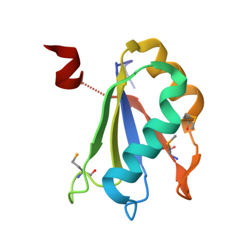
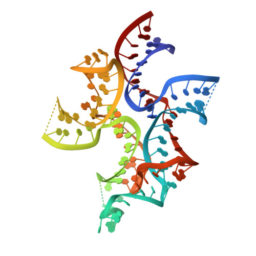
    Cyclic diadenosine monophosphate (c-di-AMP) is a second messenger that is essential for growth and homeostasis in bacteria. A recently discovered c-di-AMP-responsive riboswitch controls the expression of genes in a variety of bacteria, including important pathogens. To elucidate the molecular basis for specific binding of c-di-AMP by a gene-regulatory mRNA domain, we have determined the co-crystal structure of this riboswitch. Unexpectedly, the structure reveals an internally pseudo-symmetric RNA in which two similar three-helix-junction elements associate head-to-tail, creating a trough that cradles two c-di-AMP molecules making quasi-equivalent contacts with the riboswitch. The riboswitch selectively binds c-di-AMP and discriminates exquisitely against other cyclic dinucleotides, such as c-di-GMP and cyclic-AMP-GMP, via interactions with both the backbone and bases of its cognate second messenger. Small-angle X-ray scattering experiments indicate that global folding of the riboswitch is induced by the two bound cyclic dinucleotides, which bridge the two symmetric three-helix domains. This structural reorganization likely couples c-di-AMP binding to gene expression.


  • Organizational Affiliation

    Biochemistry and Biophysics Center, National Heart, Lung and Blood Institute, Bethesda, MD, USA.


Macromolecules

Find similar proteins by:  (by identity cutoff)  |  3D Structure
Entity ID: 1
MoleculeChains Sequence LengthOrganismDetailsImage
U1 small nuclear ribonucleoprotein AA [auth B]91Homo sapiensMutation(s): 2 
Gene Names: SNRPA
UniProt & NIH Common Fund Data Resources
Find proteins for P09012 (Homo sapiens)
Explore P09012 
Go to UniProtKB:  P09012
PHAROS:  P09012
GTEx:  ENSG00000077312 
Entity Groups  
Sequence Clusters30% Identity50% Identity70% Identity90% Identity95% Identity100% Identity
UniProt GroupP09012
Sequence Annotations
Expand
  • Reference Sequence
Find similar nucleic acids by:  (by identity cutoff)  |  3D Structure
Entity ID: 2
MoleculeChains LengthOrganismImage
C-di-AMP ribsoswitchB [auth C]119Bacillus subtilis
Sequence Annotations
Expand
  • Reference Sequence
Small Molecules
Ligands 4 Unique
IDChains Name / Formula / InChI Key2D Diagram3D Interactions
2BA
Query on 2BA

Download Ideal Coordinates CCD File 
J [auth C],
K [auth C]
(2R,3R,3aS,5R,7aR,9R,10R,10aS,12R,14aR)-2,9-bis(6-amino-9H-purin-9-yl)octahydro-2H,7H-difuro[3,2-d:3',2'-j][1,3,7,9,2,8 ]tetraoxadiphosphacyclododecine-3,5,10,12-tetrol 5,12-dioxide
C20 H24 N10 O12 P2
PDXMFTWFFKBFIN-XPWFQUROSA-N
EDO
Query on EDO

Download Ideal Coordinates CCD File 
I [auth C]1,2-ETHANEDIOL
C2 H6 O2
LYCAIKOWRPUZTN-UHFFFAOYSA-N
K
Query on K

Download Ideal Coordinates CCD File 
H [auth C]POTASSIUM ION
K
NPYPAHLBTDXSSS-UHFFFAOYSA-N
MG
Query on MG

Download Ideal Coordinates CCD File 
C,
D [auth C],
E [auth C],
F [auth C],
G [auth C]
MAGNESIUM ION
Mg
JLVVSXFLKOJNIY-UHFFFAOYSA-N
Modified Residues  1 Unique
IDChains TypeFormula2D DiagramParent
MSE
Query on MSE
A [auth B]L-PEPTIDE LINKINGC5 H11 N O2 SeMET
Experimental Data & Validation

Experimental Data

  • Method: X-RAY DIFFRACTION
  • Resolution: 3.21 Å
  • R-Value Free: 0.289 
  • R-Value Work: 0.252 
  • R-Value Observed: 0.255 
  • Space Group: I 2 2 2
Unit Cell:
Length ( Å )Angle ( ˚ )
a = 60.281α = 90
b = 83.062β = 90
c = 233.573γ = 90
Software Package:
Software NamePurpose
PHENIXrefinement
SCALEPACKdata scaling
PDB_EXTRACTdata extraction

Structure Validation

View Full Validation Report


Ligand Structure Quality Assessment 


Entry History 

Deposition Data

Revision History  (Full details and data files)

  • Version 1.0: 2014-10-22
    Type: Initial release
  • Version 1.1: 2014-10-29
    Changes: Database references
  • Version 1.2: 2014-12-03
    Changes: Database references
  • Version 1.3: 2017-11-22
    Changes: Database references, Derived calculations, Other, Refinement description, Source and taxonomy
  • Version 1.4: 2023-12-27
    Changes: Data collection, Database references, Derived calculations, Refinement description, Structure summary