4UF0

Crystal structure of JmjC domain of human histone demethylase UTY in complex with epitherapuetic compound 2-(((2-((2-(dimethylamino)ethyl) (ethyl)amino)-2-oxoethyl)amino)methyl)isonicotinic acid.


Experimental Data Snapshot

  • Method: X-RAY DIFFRACTION
  • Resolution: 1.78 Å
  • R-Value Free: 0.215 
  • R-Value Work: 0.149 
  • R-Value Observed: 0.152 

wwPDB Validation   3D Report Full Report


Ligand Structure Quality Assessment 


This is version 1.6 of the entry. See complete history


Literature

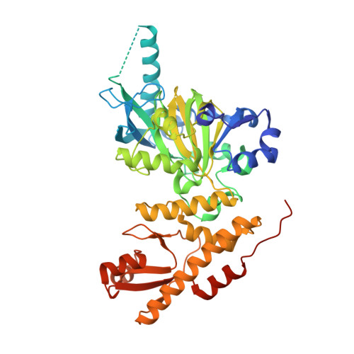
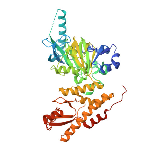
Structural Analysis of Human Kdm5B Guides Histone Demethylase Inhibitor Development.

Johansson, C.Velupillai, S.Tumber, A.Szykowska, A.Hookway, E.S.Nowak, R.P.Strain-Damerell, C.Gileadi, C.Philpott, M.Burgess-Brown, N.Wu, N.Kopec, J.Nuzzi, A.Steuber, H.Egner, U.Badock, V.Munro, S.Lathangue, N.B.Westaway, S.Brown, J.Athanasou, N.Prinjha, R.Brennan, P.E.Oppermann, U.

(2016) Nat Chem Biol 12: 539

  • DOI: https://doi.org/10.1038/nchembio.2087
  • Primary Citation of Related Structures:  
    4UF0, 5A1F, 5A3P, 5A3T, 5A3W, 5FPU, 5FPV, 5FUN, 5FUP, 5FV3, 5FWJ

  • PubMed Abstract: 

    Members of the KDM5 (also known as JARID1) family are 2-oxoglutarate- and Fe(2+)-dependent oxygenases that act as histone H3K4 demethylases, thereby regulating cell proliferation and stem cell self-renewal and differentiation. Here we report crystal structures of the catalytic core of the human KDM5B enzyme in complex with three inhibitor chemotypes. These scaffolds exploit several aspects of the KDM5 active site, and their selectivity profiles reflect their hybrid features with respect to the KDM4 and KDM6 families. Whereas GSK-J1, a previously identified KDM6 inhibitor, showed about sevenfold less inhibitory activity toward KDM5B than toward KDM6 proteins, KDM5-C49 displayed 25-100-fold selectivity between KDM5B and KDM6B. The cell-permeable derivative KDM5-C70 had an antiproliferative effect in myeloma cells, leading to genome-wide elevation of H3K4me3 levels. The selective inhibitor GSK467 exploited unique binding modes, but it lacked cellular potency in the myeloma system. Taken together, these structural leads deliver multiple starting points for further rational and selective inhibitor design.


  • Organizational Affiliation

    Structural Genomics Consortium, University of Oxford, Headington, UK.


Macromolecules
Find similar proteins by:  (by identity cutoff)  |  3D Structure
Entity ID: 1
MoleculeChains Sequence LengthOrganismDetailsImage
HISTONE DEMETHYLASE UTY478Homo sapiensMutation(s): 0 
EC: 1.14.11
UniProt & NIH Common Fund Data Resources
Find proteins for O14607 (Homo sapiens)
Explore O14607 
Go to UniProtKB:  O14607
PHAROS:  O14607
GTEx:  ENSG00000183878 
Entity Groups  
Sequence Clusters30% Identity50% Identity70% Identity90% Identity95% Identity100% Identity
UniProt GroupO14607
Sequence Annotations
Expand
  • Reference Sequence
Find similar proteins by:  (by identity cutoff)  |  3D Structure
Entity ID: 2
MoleculeChains Sequence LengthOrganismDetailsImage
HISTONE DEMETHYLASE UTY478Homo sapiensMutation(s): 0 
EC: 1.14.11
UniProt & NIH Common Fund Data Resources
Find proteins for O14607 (Homo sapiens)
Explore O14607 
Go to UniProtKB:  O14607
PHAROS:  O14607
GTEx:  ENSG00000183878 
Entity Groups  
Sequence Clusters30% Identity50% Identity70% Identity90% Identity95% Identity100% Identity
UniProt GroupO14607
Sequence Annotations
Expand
  • Reference Sequence
Small Molecules
Ligands 4 Unique
IDChains Name / Formula / InChI Key2D Diagram3D Interactions
MMK
Query on MMK

Download Ideal Coordinates CCD File 
D [auth A],
O [auth B]
2-{[(2-{[(E)-2-(dimethylamino)ethenyl](ethyl)amino}-2-oxoethyl)amino]methyl}pyridine-4-carboxylic acid
C15 H22 N4 O3
RTKGUAPXWDCFNW-BQYQJAHWSA-N
ZN
Query on ZN

Download Ideal Coordinates CCD File 
E [auth A],
P [auth B]
ZINC ION
Zn
PTFCDOFLOPIGGS-UHFFFAOYSA-N
EDO
Query on EDO

Download Ideal Coordinates CCD File 
AA [auth B]
BA [auth B]
F [auth A]
G [auth A]
H [auth A]
AA [auth B],
BA [auth B],
F [auth A],
G [auth A],
H [auth A],
I [auth A],
J [auth A],
K [auth A],
L [auth A],
M [auth A],
Q [auth B],
R [auth B],
S [auth B],
T [auth B],
U [auth B],
V [auth B],
W [auth B],
X [auth B],
Y [auth B],
Z [auth B]
1,2-ETHANEDIOL
C2 H6 O2
LYCAIKOWRPUZTN-UHFFFAOYSA-N
FE2
Query on FE2

Download Ideal Coordinates CCD File 
C [auth A],
N [auth B]
FE (II) ION
Fe
CWYNVVGOOAEACU-UHFFFAOYSA-N
Experimental Data & Validation

Experimental Data

  • Method: X-RAY DIFFRACTION
  • Resolution: 1.78 Å
  • R-Value Free: 0.215 
  • R-Value Work: 0.149 
  • R-Value Observed: 0.152 
  • Space Group: P 21 21 21
Unit Cell:
Length ( Å )Angle ( ˚ )
a = 90.494α = 90
b = 110.458β = 90
c = 119.23γ = 90
Software Package:
Software NamePurpose
REFMACrefinement
XDSdata reduction
SCALAdata scaling
CCP4Iphasing

Structure Validation

View Full Validation Report


Ligand Structure Quality Assessment 


Entry History 

Deposition Data

Revision History  (Full details and data files)

  • Version 1.0: 2015-01-14
    Type: Initial release
  • Version 1.1: 2015-07-01
    Changes: Atomic model, Database references, Derived calculations, Non-polymer description, Other
  • Version 1.2: 2016-05-25
    Changes: Database references
  • Version 1.3: 2016-06-01
    Changes: Database references
  • Version 1.4: 2016-06-29
    Changes: Database references
  • Version 1.5: 2018-01-24
    Changes: Advisory, Structure summary
  • Version 1.6: 2023-12-20
    Changes: Advisory, Data collection, Database references, Derived calculations, Other, Refinement description