4N59

The Crystal Structure of Pectocin M2 at 2.3 Angstroms


Experimental Data Snapshot

  • Method: X-RAY DIFFRACTION
  • Resolution: 2.30 Å
  • R-Value Free: 0.274 
  • R-Value Work: 0.206 
  • R-Value Observed: 0.210 

wwPDB Validation   3D Report Full Report


This is version 1.2 of the entry. See complete history


Literature

Structure of the atypical bacteriocin pectocin M2 implies a novel mechanism of protein uptake.

Grinter, R.Josts, I.Zeth, K.Roszak, A.W.McCaughey, L.C.Cogdell, R.J.Milner, J.J.Kelly, S.M.Byron, O.Walker, D.

(2014) Mol Microbiol 93: 234-246

  • DOI: https://doi.org/10.1111/mmi.12655
  • Primary Citation of Related Structures:  
    4N58, 4N59

  • PubMed Abstract: 

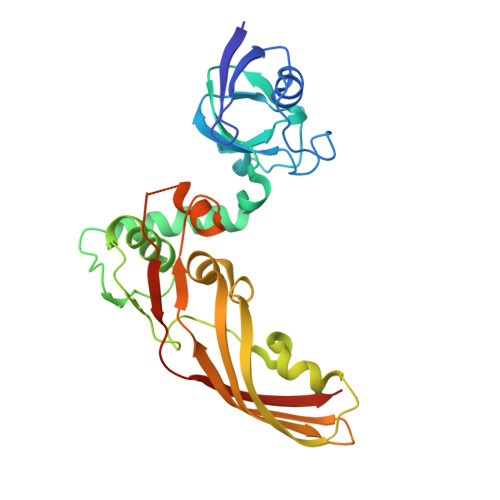
    The colicin-like bacteriocins are potent protein antibiotics that have evolved to efficiently cross the outer membrane of Gram-negative bacteria by parasitizing nutrient uptake systems. We have structurally characterized the colicin M-like bacteriocin, pectocin M2, which is active against strains of Pectobacterium spp. This unusual bacteriocin lacks the intrinsically unstructured translocation domain that usually mediates translocation of these bacteriocins across the outer membrane, containing only a single globular ferredoxin domain connected to its cytotoxic domain by a flexible α-helix, which allows it to adopt two distinct conformations in solution. The ferredoxin domain of pectocin M2 is homologous to plant ferredoxins and allows pectocin M2 to parasitize a system utilized by Pectobacterium to obtain iron during infection of plants. Furthermore, we identify a novel ferredoxin-containing bacteriocin pectocin P, which possesses a cytotoxic domain homologous to lysozyme, illustrating that the ferredoxin domain acts as a generic delivery module for cytotoxic domains in Pectobacterium.


  • Organizational Affiliation

    Institute of Infection, Immunity and Inflammation, University of Glasgow, Glasgow, G12 8QQ, UK.


Macromolecules
Find similar proteins by:  (by identity cutoff)  |  3D Structure
Entity ID: 1
MoleculeChains Sequence LengthOrganismDetailsImage
Pectocin M2
A, B
279Pectobacterium brasiliense PBR1692Mutation(s): 0 
UniProt
Find proteins for A0A067XG75 (Pectobacterium brasiliense PBR1692)
Explore A0A067XG75 
Go to UniProtKB:  A0A067XG75
Entity Groups  
Sequence Clusters30% Identity50% Identity70% Identity90% Identity95% Identity100% Identity
UniProt GroupA0A067XG75
Sequence Annotations
Expand
  • Reference Sequence
Small Molecules
Experimental Data & Validation

Experimental Data

  • Method: X-RAY DIFFRACTION
  • Resolution: 2.30 Å
  • R-Value Free: 0.274 
  • R-Value Work: 0.206 
  • R-Value Observed: 0.210 
  • Space Group: P 1 21 1
Unit Cell:
Length ( Å )Angle ( ˚ )
a = 44.649α = 90
b = 116.745β = 94.96
c = 60.79γ = 90
Software Package:
Software NamePurpose
XDSdata scaling
PHASERphasing
REFMACrefinement
XDSdata reduction

Structure Validation

View Full Validation Report


Entry History 

Deposition Data

Revision History  (Full details and data files)

  • Version 1.0: 2014-06-04
    Type: Initial release
  • Version 1.1: 2014-08-13
    Changes: Database references
  • Version 1.2: 2023-09-20
    Changes: Data collection, Database references, Derived calculations, Refinement description