4LNL

Structure of Escherichia coli Threonine Aldolase in Complex with Allo-Thr


Experimental Data Snapshot

  • Method: X-RAY DIFFRACTION
  • Resolution: 2.10 Å
  • R-Value Free: 0.243 
  • R-Value Work: 0.182 
  • R-Value Observed: 0.182 

wwPDB Validation   3D Report Full Report


Ligand Structure Quality Assessment 


This is version 1.2 of the entry. See complete history


Literature

On the catalytic mechanism and stereospecificity of Escherichia coli l-threonine aldolase.

di Salvo, M.L.Remesh, S.G.Vivoli, M.Ghatge, M.S.Paiardini, A.D'Aguanno, S.Safo, M.K.Contestabile, R.

(2014) FEBS J 281: 129-145

  • DOI: https://doi.org/10.1111/febs.12581
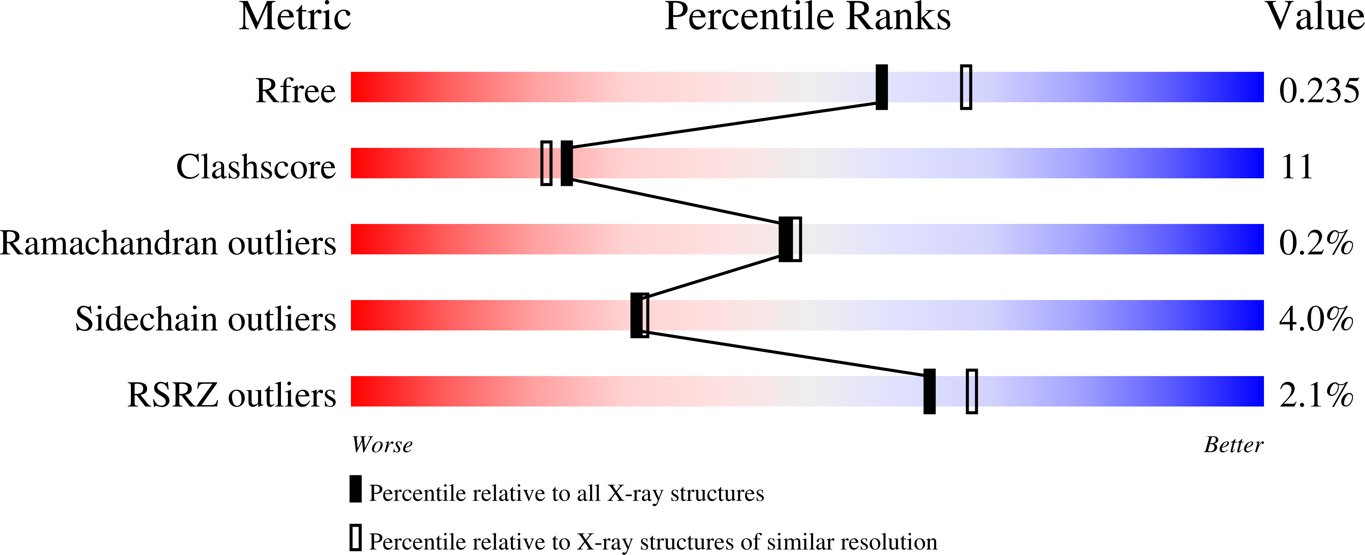
  • Primary Citation of Related Structures:  
    4LNJ, 4LNL, 4LNM

  • PubMed Abstract: 

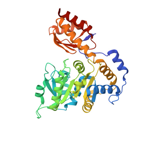
    L-threonine aldolases (L-TAs) represent a family of homologous pyridoxal 5'-phosphate-dependent enzymes found in bacteria and fungi, and catalyse the reversible cleavage of several L-3-hydroxy-α-amino acids. L-TAs have great biotechnological potential, as they catalyse the formation of carbon-carbon bonds, and therefore may be exploited for the bioorganic synthesis of L-3-hydroxyamino acids that are biologically active or constitute building blocks for pharmaceutical molecules. Many L-TAs, showing different stereospecificity towards the Cβ configuration, have been isolated. Because of their potential to carry out diastereoselective syntheses, L-TAs have been subjected to structural, functional and mechanistic studies. Nevertheless, their catalytic mechanism and the structural bases of their stereospecificity have not been elucidated. In this study, we have determined the crystal structure of low-specificity L-TA from Escherichia coli at 2.2-Å resolution, in the unliganded form and cocrystallized with L-serine and L-threonine. Furthermore, several active site mutants have been functionally characterized in order to elucidate the reaction mechanism and the molecular bases of stereospecificity. No active site catalytic residue was revealed, and a structural water molecule was assumed to act as the catalytic base in the retro-aldol cleavage reaction. Interestingly, the very large active site opening of E. coli L-TA suggests that much larger molecules than L-threonine isomers may be easily accommodated, and L-TAs may actually have diverse physiological functions in different organisms. Substrate recognition and reaction specificity seem to be guided by the overall microenvironment that surrounds the substrate at the enzyme active site, rather than by one ore more specific residues.


  • Organizational Affiliation

    Dipartimento di Scienze Biochimiche 'A. Rossi Fanelli', Sapienza Università di Roma, Italy.


Macromolecules
Find similar proteins by:  (by identity cutoff)  |  3D Structure
Entity ID: 1
MoleculeChains Sequence LengthOrganismDetailsImage
Low-specificity L-threonine aldolase
A, B
333Escherichia coli WV_060327Mutation(s): 0 
Gene Names: EcoM_01176ltaE
EC: 4.1.2.5
Entity Groups  
Sequence Clusters30% Identity50% Identity70% Identity90% Identity95% Identity100% Identity
Sequence Annotations
Expand
  • Reference Sequence
Small Molecules
Ligands 6 Unique
IDChains Name / Formula / InChI Key2D Diagram3D Interactions
2BK
Query on 2BK

Download Ideal Coordinates CCD File 
E [auth A]N-({3-hydroxy-2-methyl-5-[(phosphonooxy)methyl]pyridin-4-yl}methyl)-L-allothreonine
C12 H19 N2 O8 P
IZWQBQLGLAKRMN-XVKPBYJWSA-N
2BO
Query on 2BO

Download Ideal Coordinates CCD File 
D [auth A]N-({3-hydroxy-2-methyl-5-[(phosphonooxy)methyl]pyridin-4-yl}methyl)-L-threonine
C12 H19 N2 O8 P
IZWQBQLGLAKRMN-XCBNKYQSSA-N
PLG
Query on PLG

Download Ideal Coordinates CCD File 
J [auth B]N-GLYCINE-[3-HYDROXY-2-METHYL-5-PHOSPHONOOXYMETHYL-PYRIDIN-4-YL-METHANE]
C10 H15 N2 O7 P
FEVQWBMNLWUBTF-UHFFFAOYSA-N
EPE
Query on EPE

Download Ideal Coordinates CCD File 
F [auth A],
K [auth B]
4-(2-HYDROXYETHYL)-1-PIPERAZINE ETHANESULFONIC ACID
C8 H18 N2 O4 S
JKMHFZQWWAIEOD-UHFFFAOYSA-N
CL
Query on CL

Download Ideal Coordinates CCD File 
G [auth A]CHLORIDE ION
Cl
VEXZGXHMUGYJMC-UHFFFAOYSA-M
MG
Query on MG

Download Ideal Coordinates CCD File 
C [auth A],
H [auth B],
I [auth B]
MAGNESIUM ION
Mg
JLVVSXFLKOJNIY-UHFFFAOYSA-N
Experimental Data & Validation

Experimental Data

  • Method: X-RAY DIFFRACTION
  • Resolution: 2.10 Å
  • R-Value Free: 0.243 
  • R-Value Work: 0.182 
  • R-Value Observed: 0.182 
  • Space Group: C 2 2 21
Unit Cell:
Length ( Å )Angle ( ˚ )
a = 76.4α = 90
b = 100.89β = 90
c = 176γ = 90
Software Package:
Software NamePurpose
CNSrefinement
CrystalCleardata collection
d*TREKdata reduction
d*TREKdata scaling
CaspRphasing

Structure Validation

View Full Validation Report


Ligand Structure Quality Assessment 


Entry History 

Deposition Data

  • Released Date: 2013-11-06 
  • Deposition Author(s): Safo, M.K.

Revision History  (Full details and data files)

  • Version 1.0: 2013-11-06
    Type: Initial release
  • Version 1.1: 2014-01-22
    Changes: Database references
  • Version 1.2: 2023-09-20
    Changes: Data collection, Database references, Derived calculations, Refinement description