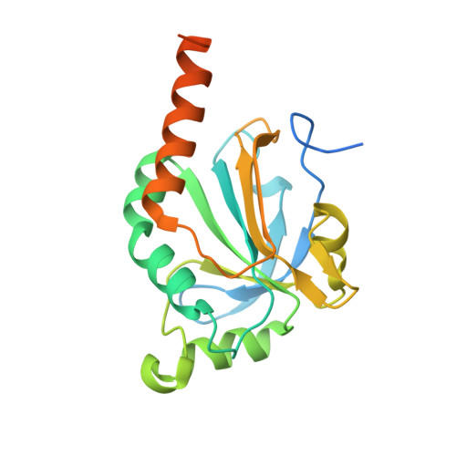
Observed octameric assembly of a Plasmodium yoelii peroxiredoxin can be explained by the replacement of native "ball-and-socket" interacting residues by an affinity tag.
Gretes, M.C., Karplus, P.A.(2013) Protein Sci 22: 1445-1452
- PubMed: 23934758
- DOI: https://doi.org/10.1002/pro.2328
- Primary Citation of Related Structures:
4L0U, 4L0W - PubMed Abstract:
Peroxiredoxins (Prxs) are ubiquitous and efficient antioxidant enzymes crucial for redox homeostasis in most organisms, and are of special importance for disease-causing parasites that must protect themselves against the oxidative weapons of the human immune system. Here, we describe reanalyses of crystal structures of two Prxs from malaria parasites. In addition to producing improved structures, we provide normalizing explanations for features that had been noted as unusual in the original report of these structures (Qiu et al., BMC Struct Biol 2012;12:2). Most importantly, we provide evidence that the unusual octameric assembly seen for Plasmodium yoelii Prx1a is not physiologically relevant, but arises because the structure is not of authentic P. yoelii Prx1a, but a variant we designate PyPrx1a(N*) that has seven native N-terminal residues replaced by an affinity tag. This N-terminal modification disrupts a previously unrecognized, hydrophobic "ball-and-socket" interaction conserved at the B-type dimer interface of Prx1 subfamily enzymes, and is accommodated by a fascinating two-residue "β-slip" type register shift in the β-strand association at a dimer interface. The resulting change in the geometry of the dimer provides a simple explanation for octamer formation. This study illustrates how substantive impacts can occur in protein variants in which native residues have been altered.
Organizational Affiliation:
Department of Biochemistry & Molecular Biology, Oregon Health & Science University, Portland, Oregon, 97239; Department of Biochemistry & Biophysics, Oregon State University, Corvallis, Oregon, 97331.