4IQH

Crystal Structure Analysis of Dysferlin C2A variant 1 (C2Av1)


Experimental Data Snapshot

  • Method: X-RAY DIFFRACTION
  • Resolution: 1.76 Å
  • R-Value Free: 0.215 
  • R-Value Work: 0.192 
  • R-Value Observed: 0.195 

wwPDB Validation   3D Report Full Report


This is version 1.3 of the entry. See complete history


Literature

Alternate Splicing of Dysferlin C2A Confers Ca(2+)-Dependent and Ca(2+)-Independent Binding for Membrane Repair.

Fuson, K.Rice, A.Mahling, R.Snow, A.Nayak, K.Shanbhogue, P.Meyer, A.G.Redpath, G.M.Hinderliter, A.Cooper, S.T.Sutton, R.B.

(2014) Structure 22: 104-115

  • DOI: https://doi.org/10.1016/j.str.2013.10.001
  • Primary Citation of Related Structures:  
    4IHB, 4IQH

  • PubMed Abstract: 

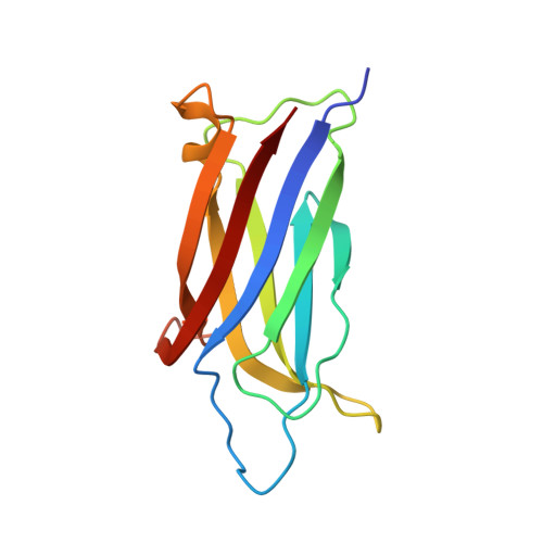
    Dysferlin plays a critical role in the Ca²⁺-dependent repair of microlesions that occur in the muscle sarcolemma. Of the seven C2 domains in dysferlin, only C2A is reported to bind both Ca²⁺ and phospholipid, thus acting as a key sensor in membrane repair. Dysferlin C2A exists as two isoforms, the "canonical" C2A and C2A variant 1 (C2Av1). Interestingly, these isoforms have markedly different responses to Ca²⁺ and phospholipid. Structural and thermodynamic analyses are consistent with the canonical C2A domain as a Ca²⁺-dependent, phospholipid-binding domain, whereas C2Av1 would likely be Ca²⁺-independent under physiological conditions. Additionally, both isoforms display remarkably low free energies of stability, indicative of a highly flexible structure. The inverted ligand preference and flexibility for both C2A isoforms suggest the capability for both constitutive and Ca²⁺-regulated effector interactions, an activity that would be essential in its role as a mediator of membrane repair.


  • Organizational Affiliation

    Department of Cell Physiology and Molecular Biophysics, Texas Tech University Health Sciences Center, Lubbock, TX 79430, USA; Center for Membrane Protein Research, Texas Tech University Health Sciences Center, Lubbock, TX 79430, USA.


Macromolecules
Find similar proteins by:  (by identity cutoff)  |  3D Structure
Entity ID: 1
MoleculeChains Sequence LengthOrganismDetailsImage
Dysferlin
A, B, C
130Homo sapiensMutation(s): 0 
Gene Names: DYSFDysferlinFER1L1
UniProt & NIH Common Fund Data Resources
Find proteins for O75923 (Homo sapiens)
Explore O75923 
Go to UniProtKB:  O75923
PHAROS:  O75923
GTEx:  ENSG00000135636 
Entity Groups  
Sequence Clusters30% Identity50% Identity70% Identity90% Identity95% Identity100% Identity
UniProt GroupO75923
Sequence Annotations
Expand
  • Reference Sequence
Experimental Data & Validation

Experimental Data

  • Method: X-RAY DIFFRACTION
  • Resolution: 1.76 Å
  • R-Value Free: 0.215 
  • R-Value Work: 0.192 
  • R-Value Observed: 0.195 
  • Space Group: P 31 2 1
Unit Cell:
Length ( Å )Angle ( ˚ )
a = 71.16α = 90
b = 71.16β = 90
c = 137.45γ = 120
Software Package:
Software NamePurpose
HKL-3000data collection
PHASERphasing
PHENIXrefinement
HKL-3000data reduction
SCALEPACKdata scaling

Structure Validation

View Full Validation Report


Entry History 

Deposition Data

Revision History  (Full details and data files)

  • Version 1.0: 2013-12-18
    Type: Initial release
  • Version 1.1: 2014-01-29
    Changes: Database references
  • Version 1.2: 2018-04-25
    Changes: Data collection
  • Version 1.3: 2023-09-20
    Changes: Data collection, Database references, Refinement description