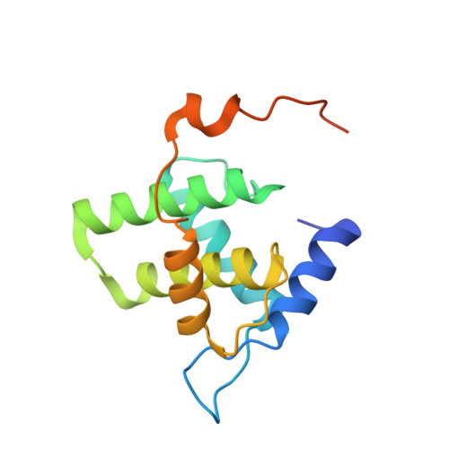
Biophysical characterization and crystal structure of the Feline Immunodeficiency Virus p15 matrix protein.
Serriere, J., Robert, X., Perez, M., Gouet, P., Guillon, C.(2013) Retrovirology 10: 64-64
- PubMed: 23800358
- DOI: https://doi.org/10.1186/1742-4690-10-64
- Primary Citation of Related Structures:
4IC9, 4ICA - PubMed Abstract:
Feline Immunodeficiency Virus (FIV) is a viral pathogen that infects domestic cats and wild felids. During the viral replication cycle, the FIV p15 matrix protein oligomerizes to form a closed matrix that underlies the lipidic envelope of the virion. Because of its crucial role in the early and late stages of viral morphogenesis, especially in viral assembly, FIV p15 is an interesting target in the development of potential new therapeutic strategies. Our biochemical study of FIV p15 revealed that it forms a stable dimer in solution under acidic conditions and at high concentration, unlike other retroviral matrix proteins. We determined the crystal structure of full-length FIV p15 to 2 Å resolution and observed a helical organization of the protein, typical for retroviral matrix proteins. A hydrophobic pocket that could accommodate a myristoyl group was identified, and the C-terminal end of FIV p15, which is mainly unstructured, was visible in electron density maps. As FIV p15 crystallizes in acidic conditions but with one monomer in the asymmetric unit, we searched for the presence of a biological dimer in the crystal. No biological assembly was detected by the PISA server, but the three most buried crystallographic interfaces have interesting features: the first one displays a highly conserved tryptophan acting as a binding platform, the second one is located along a 2-fold symmetry axis and the third one resembles the dimeric interface of EIAV p15. Because the C-terminal end of p15 is involved in two of these three interfaces, we investigated the structure and assembly of a C-terminal-truncated form of p15 lacking 14 residues. The truncated FIV p15 dimerizes in solution at a lower concentration and crystallizes with two molecules in the asymmetric unit. The EIAV-like dimeric interface is the only one to be retained in the new crystal form. The dimeric form of FIV p15 in solution and its extended C-terminal end are characteristic among lentiviral matrix proteins. Crystallographic interfaces revealed several interactions that might be involved in FIV replication. Further studies are needed to better understand their biological relevance in the function of FIV Gag during viral replication.