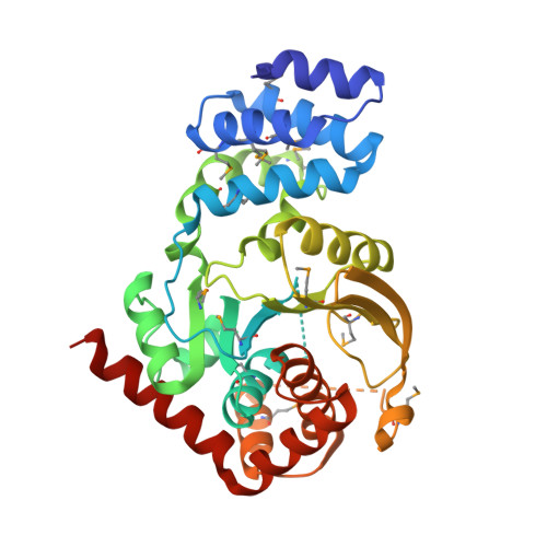
Crystal Structure of an Anthranilate Phosphoribosyltransferase (target ID NYSGRC-016600) from Xanthomonas campestris
Ghosh, A., Ahmed, A., Banu, R., Bhoshle, R., Bonanno, J., Chamala, S., Chowdhury, S., Fiser, A., Glenn, A.S., Hillerich, B., Khafizov, K., Lafleur, J., Love, J.D., Seidel, R., Stead, M., Toro, R., Almo, S.C.To be published.